
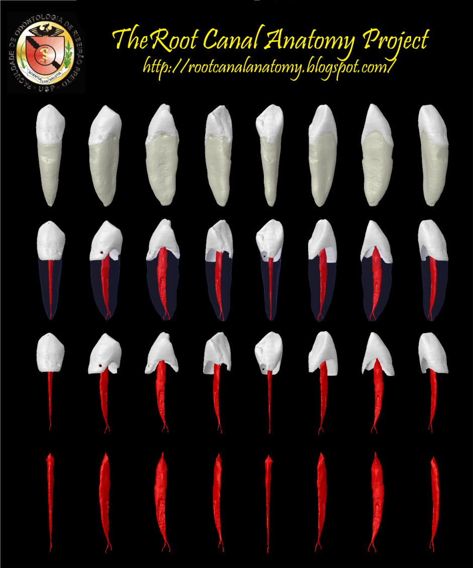
Sturdy and considerably wider mesial-distally than the incisors, the mandibular canines seldom present endodontic problems. The unusual occurrence of two roots can create difficulty, but this is rare. The access cavity is ovoid and may be extended incisally for labial-lingual accessibility. The canal is somewhat ovoid at the cervical, becoming round at midroot. Directional instrumentation is necessary to debride the canal walls completely. If there are two roots, one is always easier to instrument. The other must be opened and funneled in concert with the first to prevent packing of dentin debris and loss of access. Precurving of instruments at initial access will enable the clinician to trace down the buccal or lingual root wall until the tip engages the orifice. When the difficult canal is located, every effort should be made to shape and funnel the opening to maintain continued access (Burns RC, Buchanan LS. Tooth Morphology and Access Openings. Part One: The Art of Endodontics in Pathway of Pulp, 6th Ed. p. 154).

Keywords: micro-computed tomography, micro-ct, marco versiani, micro-computer tomography, high resolution x-ray tomography, dental anatomy, root canal anatomy
No comments:
Post a Comment