From 2011-2016, images and videos of "The Root Canal Anatomy Project" were developed at the Laboratory of Endodontics of Ribeirao Preto Dental School. From 2016, images were acquired in other educational institutions. They can be freely used for attributed noncommercial educational purposes by educators, scholars, student and clinicians. It means that all material used should include proper attribution and citation (http://rootcanalanatomy.blogspot.com). In such cases, this information should be linked to the image in a manner compatible with such instructional objectives. Unfortunately, because material shared on the RCAP has not been properly cited by several users, from November 2019 a watermark was added to the images and videos. Enjoy!

December 12, 2015

Publication: Comparative accuracy of the Clearing Technique, CBCT and Micro-CT

COMPARATIVE ACCURACY OF THE CLEARING TECHNIQUE, CBCT, AND MICRO-CT METHODS IN STUDYING MESIAL ROOT CANAL CONFIGURATION OF MANDIBULAR FIRST MOLARS


R Ordinola-Zapata, C M. Bramante, M A. Versiani, I B Moldauer, G Topham, 
J L. Gutmann, A Nuñez, M A Hungaro Duarte and F Abella

Despite the considerable number of studies published on the internal anatomy of posterior teeth, very little information exists regarding the accuracy of clearing, CBCT, and micro-CT methods to diagnose the morphology of the root canal anatomy. Therefore, the aim of this study was to compare the accuracy of the clearing technique and CBCT scanning in the assessment of the mesial root canal configuration of mandibular first molars, using micro-CT imaging system as a reference standard. 


November 29, 2015

Peg-Shaped Lateral Incisors (Conoid)

Peg-Shaped Lateral Incisors

The most common anomaly in tooth shape in the anterior of the secondary dentition is the peg-shaped (or cone-shaped) lateral incisor, occurring in 1-2% of the population. The tooth is generally conical in shape, broadest cervically, and tapers toward the incisal to a blunt point. Several studies of the identical twins seem to indicate that missing and peg-shaped lateral incisor teeth may be varied expressions of the same generic trait. A most unusual occurrence is that of peg-shaped maxillary central incisor. Peg-shaped teeth develop from one facial lobe (instead of the three facial lobes normally present on anterior teeth).

Woelfel & Scheid. Dental Anatomy: Its Relevance to Dentistry. 6th Ed. 2002.page 331


November 27, 2015

Publication: Review on Dentinal Microcracks - ETopics


Abstract

The dentinal radicular microcrack is a difficult clinical problem to diagnose and treat, and it is one of the most common reasons for tooth extraction. These microcracks start in the radicular dentin, and laboratory studies have linked crack formation to some routine endodontic procedures, namely root canal preparation, obturation, and retreatment. Most of these studies were performed using destructive methods, such as the sectioning technique, previously developed for the study of the internal anatomy of teeth. Nowadays, technological advances in the field of imaging may lead to a more thorough understanding of dentinal microcracks. This article seeks to critically appraise the methodological aspects involved in the study of dentinal radicular microcrack formation after root canal preparation, obturation, and retreatment.

For more information please click HERE or HERE


November 12, 2015

Book Chapter: The Root Canal Biofilm

Very honored for being invited by Dr Luis E. Chávez de Paz, Dr. Christine M. Sedgley and Dr. Anil Kishen to write the chapter entitled "Root Canal Anatomy: Implications in Biofilm Disinfection" (pages 155-187) with Dr. Ronald Ordinola-Zapata in their new book "The Root Canal Biofilm". For more information, please click HERE.


November 8, 2015

Publication: Middle Mesial Canal

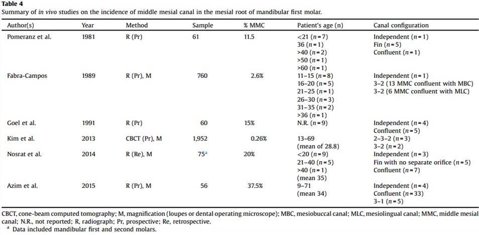
Middle mesial canals in mandibular first molars: 
a micro-CT study in different populations

Marco AurĂ©lio Versiani, Ronald Ordinola-Zapata, Ali KeleĹź, Hatice Alcin, 
ClĂłvis Monteiro Bramante, Jesus Djalma PĂ©cora, Manoel DamiĂŁo Sousa-Neto

Objective
To describe the morphological aspects of middle mesial canals (MMC) in mandibular first molars using micro-CT.

Design
Mandibular first molars collected from the Brazilian (n=136) and Turkish (n=122) populations were scanned (voxel size: 9.9 μm) and mesial roots with MMC (n=48) evaluated regarding several morphological aspects. The incidence of MMC in each population was statistically compared using Chi-square test (α=0.05).

Results
Overall, the incidence of MMC was 18.6% (48 out of 258 molars) and was significantly higher in the Brazilian (n=30; 22.1%) than in the Turkish (n=18; 14.8%) population (p<0.05). In both populations, confluent configuration of the MMC was the most frequent anatomy. Most of the specimens with MMC had 3 independent orifices (n=26; 54.2%) and 3 apical foramina (n=21; 43.8%). The mean minor diameter of the MMC orifice (0.16 mm) was 3 times less than the other orifices (∼0.50 mm). In mesial roots with independent configuration (n=3; 6.3%), the mean volumes (mm3) of the MMC, mesiobuccal (MBC) and mesiolingual (MLC) canals were 0.20±0.10, 0.75±0.28, and 0.88±0.19, respectively. In the specimens with canal confluence (n=26; 54.2%), MMC merged to the MBC (n=8; 16.7%), MLC (n=4; 8.3%), or to both MBC and MLC (n=14; 29.2%). Double mesial canal was observed in only 1 specimen. MMC with an independent foramen was observed mostly in Brazilian specimens.

Conclusions
Incidence of MMC was higher in the Brazilian molars. Confluent configuration was the most revalent anatomic variation, while independent and fin configurations, as well as, double MMC, were found only in a few specimens.

Keywords
mandibular molar; middle mesial canal; micro-CT; root canal anatomy

Click HERE or HERE to read and download the article in full


November 5, 2015

Publication: International Endodontic Journal

Micro-CT evaluation of the efficacy of hard tissue removal from the root canal and isthmus area by positive and negative pressure irrigation systems

M. A. Versiani, F. R. F. Alves, C. V. Andrade-Junior, M. F. Marceliano-Alves, J. C. Provenzano, I.N.Rocas, M. D. Sousa-Neto & J. F. Siqueira Jr


October 31, 2015

Two-Rooted Maxillary Central Incisor

A review of literature reveals that the anatomy of maxillary central incisors is single-rooted teeth with a single canal in 100% of cases. The prevalence of maxillary central incisors with 2 roots is extremely rare and has never been investigated; only a few clinical case reports have been published (Levin et al. 2015). 


Below there is a similar case report from Levin et al. published in the Journal of Endodontics
This article can be read in full HERE