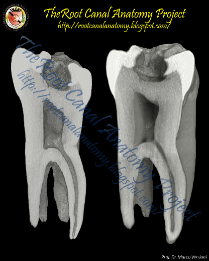
From 2011-2016, images and videos of "The Root Canal Anatomy Project" were developed at the Laboratory of Endodontics of Ribeirao Preto Dental School. From 2016, images were acquired in other educational institutions. They can be freely used for attributed noncommercial educational purposes by educators, scholars, student and clinicians. It means that all material used should include proper attribution and citation (http://rootcanalanatomy.blogspot.com). In such cases, this information should be linked to the image in a manner compatible with such instructional objectives. Unfortunately, because material shared on the RCAP has not been properly cited by several users, from November 2019 a watermark was added to the images and videos. Enjoy!