From 2011-2016, images and videos of "The Root Canal Anatomy Project" were developed at the Laboratory of Endodontics of Ribeirao Preto Dental School. From 2016, images were acquired in other educational institutions. They can be freely used for attributed noncommercial educational purposes by educators, scholars, student and clinicians. It means that all material used should include proper attribution and citation (http://rootcanalanatomy.blogspot.com). In such cases, this information should be linked to the image in a manner compatible with such instructional objectives. Unfortunately, because material shared on the RCAP has not been properly cited by several users, from November 2019 a watermark was added to the images and videos. Enjoy!

November 16, 2014

Root Canal Preparation: Mandibular Molar

Root Canal Preparation of a Mandibular Molar 
Using a Multiple-File Rotary NiTi System

Thanks to Dr. Ali Keles from Turkey for the scanning and canal preparation procedures
In green: root canal before preparation
In red: root canal after preparation superimposed to the original canal


October 28, 2014

Primary Mandibular First Molar

Primary mandibular molars have been usually described as having two grooved and divergent roots that flare to accommodate the developing permanent premolars. In the literature, a considerable variation in number and shape of canal systems has been described in this group of teeth. Overall, it may be inferred that the external and internal anatomy of the primary mandibular first molar closely resembles the primary mandibular second molar. Most studies have found either one or two canals in each of the mesial and distal roots.


September 9, 2014

Publication: Clinical Oral Investigations

Abstract

Objectives
The aim of this study was to introduce a methodology to map irrigant spreadability within the root canal space using micro-computed tomography (micro-CT).

Materials and methods
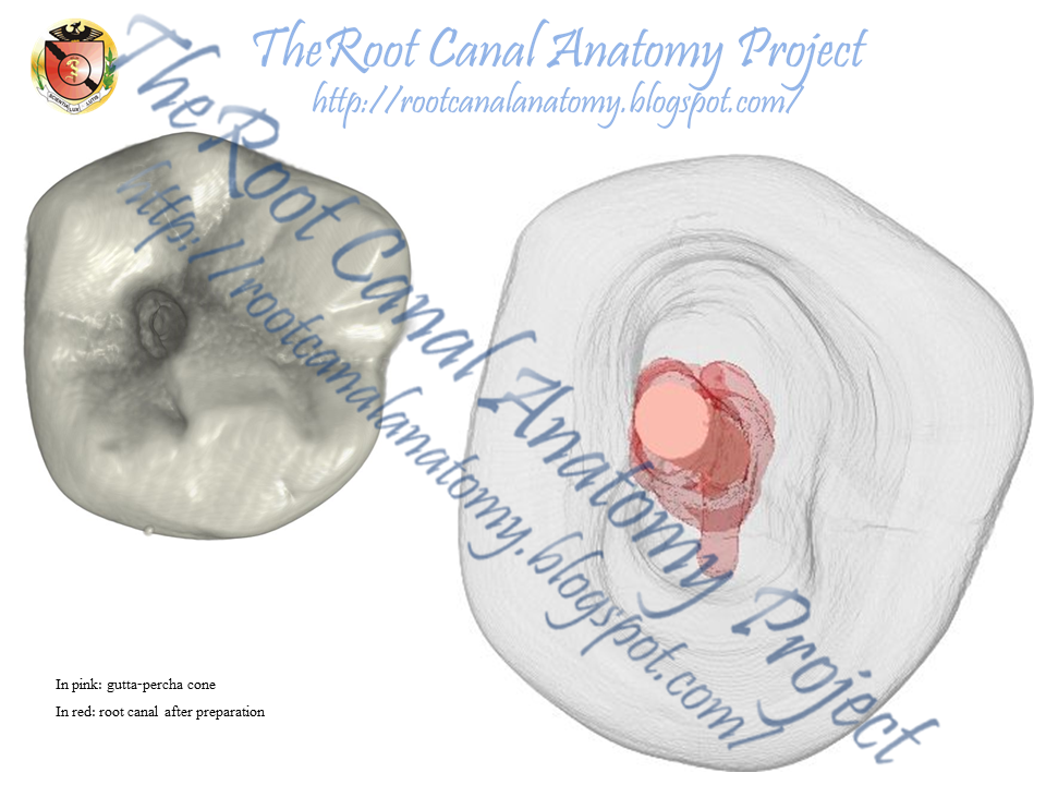
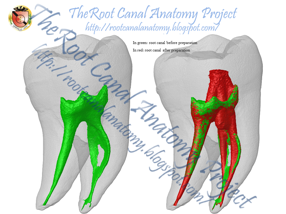
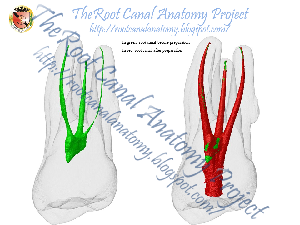
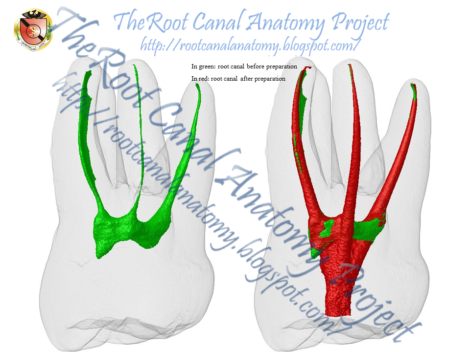
Mandibular molars presenting Vertucci’s types I and II canal configurations were selected, and four scans using isotropic resolution of 19.5 μm were accomplished per tooth: prior to treatment (S1), after glide path (S2) and after root canal preparation (S3 and S4). A contrast solution (CS) was used to irrigate the canals at stages S2 and S4. The touched and untouched surface areas of the canals, the volume of irrigant-free areas and the percentage volume occupied by the CS were calculated. Density, surface tension and the spread pattern of the CS and 2.5 % NaOCl were also evaluated.

Results
In the type I mesial root, there was an increase in the percentage volume of free-irrigated areas from S2 to S4 preparation steps, whilst in the distal roots and type II mesial root, a decrease of irrigant-free areas was observed. The use of CS allowed the quantification of the touched surface area and the volume of the root canal occupied by the irrigating solution. Density (g/mL) and surface tension (mN/m) of the CS and 2.5 % NaOCl were 1.39 and 47.5, and 1.03 and 56.2, respectively. Besides, a similar spread pattern of the CS and 2.5 % NaOCl in a simulated root canal environment was observed.

Conclusions
This study introduced a new methodology for mapping the irrigating solution in the different stages of the root canal preparation and proved useful for in situ volumetric quantification and qualitative evaluation of irrigation spreading and irrigant-free areas.

Clinical relevance
Micro-computed tomographic technology may provide a comprehensive knowledge of the flush effectiveness by different irrigants and delivery systems in order to predict the optimal cleaning and disinfection conditions of the root canal space.


August 27, 2014

Grossman's Endodontic Practice 13th edition

The current edition builds up on this platform established by the previous edition by updating and revising concepts, materials and techniques. “Clinical Notes” boxes have been introduced in each chapter highlighting the pertinent important clinical aspects of the topic being discussed for the benefit of the readers. The style and format of presentation has also been changed to make it reader friendly. Accompanying the text is a “Visual Masterclass DVD” presenting videos of important clinical procedures. Two new chapters were added: “Regenerative Endodontics” and “Endodontic Emergencies”. This book contains over 1100 figures, radiographs and illustrations; many of which are contributions from clinicians and academicians from across the world.


The Department of Endodontics of the University of Sao Paulo, Brazil, is honored for being invited to contribute with this outstanding book. Thanks to the Editors, Dr. Suresh Chandra and Dr. Gopikrishna, for this unique opportunity!

Prof. Marco Versiani
Prof. Pecora
Prof. Sousa-Neto 

August 15, 2014

Conservative Endodontic Access - Mandibular Premolar

Conservative Endodontic Access - Mandibular Premolar

The principles of traditional endodontic cavities were established many decades ago and have remained unchanged over time. The principles of traditional endodontic cavities were established many decades ago and have remained unchanged over time. The  emerging concept of conservative endodontic access prioritizes the removal of restorative materials before tooth structure, of enamel before dentin and of occlusal tooth structure before cervical dentin. It disregards the traditional requirements of a straight-line access and complete unroofing of the pulp chamber and it emphasizes the importance of preservation of the crucial pericervical dentin (located 4 mm above and below the crestal bone) to the greatest extent possible.


Unlocking the Mystery of Endodontic Treatment Success

Anatomical complexities of root canals represent the main issue in the root canal treatment. Recently, micro-CT imaging technology has been used as a powerful research tool in endodontics. This presentation will focus on the complexity of the internal anatomy of teeth and its relationship on the understanding of the principles of canal preparation and obturation. For registration click here: https://schilderinstitute.com/event/unlocking-the-mystery-of-endodontic.


Speakers and Topics

Marco Versiani
The anatomical complexity of root canal system and its relationship to endodontic treatment

Martin Trope
Biological requirements for successful endodontics

Fred Barnett
Periapical bacterial plaque in teeth refractory to endodontic treatment

John Stropko
Surgical management of cases that won’t heal

Roberto Cristescu
The access cavity - design characteristics affecting the outcome of the endodontic treatment

August 10, 2014

Conservative Endodontic Access - Mandibular Molar

Conservative Endodontic Access - Mandibular Molar

The principles of traditional endodontic cavities were established many decades ago and have remained unchanged over time. The principles of traditional endodontic cavities were established many decades ago and have remained unchanged over time. The  emerging concept of conservative endodontic access prioritizes the removal of restorative materials before tooth structure, of enamel before dentin and of occlusal tooth structure before cervical dentin. It disregards the traditional requirements of a straight-line access and complete unroofing of the pulp chamber and it emphasizes the importance of preservation of the crucial pericervical dentin (located 4 mm above and below the crestal bone) to the greatest extent possible.


August 2, 2014

Conservative Endodontic Access - Maxillary Molar

Conservative Endodontic Access - Maxillary Molar

The principles of traditional endodontic cavities were established many decades ago and have remained unchanged over time. The principles of traditional endodontic cavities were established many decades ago and have remained unchanged over time. The  emerging concept of conservative endodontic access prioritizes the removal of restorative materials before tooth structure, of enamel before dentin and of occlusal tooth structure before cervical dentin. It disregards the traditional requirements of a straight-line access and complete unroofing of the pulp chamber and it emphasizes the importance of preservation of the crucial pericervical dentin (located 4 mm above and below the crestal bone) to the greatest extent possible.