

May 24, 2014


May 19, 2014

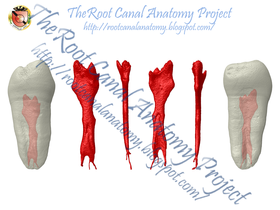
JOE Publication: Radix

Introduction: The morphology of the supernumerary third root (radix) in mandibular first molars was examined by micro–computed tomography (mCT) scanning.
Methods: Nineteen permanent mandibular first molars with radix were scanned in amCT device to evaluate their morphology with respect to root length, root curvature direction, location of radix, apical foramen, accessory canals and apical deltas, and distance between canal orifices as well as 2- and 3-dimensional parameters of the canals (number, area, roundness, major/minor diameter, volume, surface area, and structure model index). Quantitative data were analyzed by 1-way analysis of variance and the Tukey test (P<.05).
Results: The mean length of the mesial, distal, and radix roots was 20.36 ± 1.73 mm, 20.0 ± 1.83 mm, and 18.09 ± 1.68 mm, respectively. The radix was located distolingually (n= 16), mesiolingually (n= 1), and distobuccally (n= 2). In a proximal view, most radix roots had a severe curvature with buccal orientation and a buccally displaced apical foramen. The spatial configuration of the canal orifices on the pulp chamber floor was mostly in a trapezoidal shape. The radix root canal orifice was usually covered by a dentinal projection. The radix differed significantly from the mesial and distal roots for all evaluated 3-dimensional parameters (P< .05). The radix canal had a more circular shape in the apical third, and the mean size of the minor diameter 1 mm short of the foramen was 0.25 ± 0.10 mm.
Conclusions: The radix root is an important and challenging anatomic variation of mandibular first molars, which usually has a severe curvature with a predominantly distolingual location, and a narrow root canal with difficult access.


May 8, 2014
Bruker-MicroCT Meeting 2014 - Belgium
Prof. Dr. Marco Versiani - University of Sao Paulo, Brazil
5-8 May - Ostend / Belgium 2014
3D Mapping of the Irrigated Areas of the Root Canal Using Micro-CT


Prof. Dr. Graziela Bianchi Leoni - University of Sao Paulo, Brazil
5-8 May - Ostend / Belgium 2014
Push-out of root canal filling using the material testing stage (MTS)
inside a Micro-CT: preliminary observations


Subscribe to:
Posts (Atom)