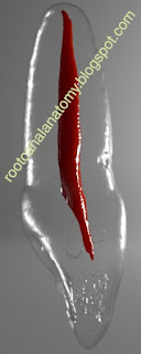
Click HERE to download the images and video in HD
From 2011-2016, images and videos of "The Root Canal Anatomy Project" were developed at the Laboratory of Endodontics of Ribeirao Preto Dental School. From 2016, images were acquired in other educational institutions. They can be freely used for attributed noncommercial educational purposes by educators, scholars, student and clinicians. It means that all material used should include proper attribution and citation (http://rootcanalanatomy.blogspot.com). In such cases, this information should be linked to the image in a manner compatible with such instructional objectives. Unfortunately, because material shared on the RCAP has not been properly cited by several users, from November 2019 a watermark was added to the images and videos. Enjoy!
December 20, 2019
December 12, 2019
MB2 canal: Systematic Review and Meta-Analysis
Summary
Prevalence studies using CBCT technology on MB2 canal were searched between May and September 2019. 83 studies were submitted to full text analysis and scientific merit evaluation by 2 evaluators and 26 studies were pooled into a meta-analysis. The included studies reported data of 23,926 maxillary molars (15,285 maxillary first molars and 8,641 maxillary second molars) from at least 12,456 patients, comprising 5,541 males and 6,915 females (2 studies did not report the number of patients). The average age of the patients was 40.9 years and was calculated based on 20 studies that reported this information. The included studies were published in English (n=24), Chinese (n=1) and Portuguese (n=1) and represented data from 24 countries.
Overall prevalence of MB2 canal
In the present study, prevalence of MB2 canal in maxillary first molars ranged from 96.7% (Belgium sub-population) to 30.9% (Chinese sub-population) while in second molars, the highest and lowest prevalence were reported in the Brazilian (83.2%) and Chinese (13.4%) sub-populations. Overall, mean prevalence of MB2 was higher in maxillary first molars (69.6%) than in second molars (39.0%). The presence of MB2 canal in maxillary first molars were addressed in 22 studies (41 population groups) with a high heterogeneity values for both maxillary first and second molars.
MB2 canal and gender
Influence of gender on the prevalence of MB2 canal in maxillary first molars was compared in 16 studies (35 population groups). Statistical comparison of untransformed proportions of MB2 for males (71.9%; 66.5%-77.4%) and females (66.8%; 60.4%-73.2%) was not significant. Meta-analysis calculation of 11 studies (12 population groups) on MB2 canal in maxillary second molars showed a high heterogeneity value and no statistical difference in its prevalence when comparing males (38.6%; 30.7%-46.5%) with females (32.1%; 23.9%-40.2%).
MB2 canal and age
The influence of age on the prevalence of the MB2 canal in maxillary first and second molars was assessed in 11 (30 population groups) and 8 (9 population groups) studies, respectively. Meta-regression calculation depicted a constant MB2 prevalence over the years and omnibus p-value excluded age as a source variance of heterogeneity.
MB2 canal and geographic region
Geographic region meta-analysis on MB2 prevalence in maxillary first and second molars were performed in 22 (41 population groups) and 16 (17 population groups) studies, respectively. In maxillary first molars, the highest proportion of MB2 canal was observed in Africa (80.9%; 67.7%-93.8%) (4 population groups combined) and the lowest in Oceania (53.1%; 46.6%-59.7%) (1 single population group), with statistical difference among a few regions. Regarding maxillary second molars, Africa showed also the highest MB2 prevalence (62.4%; 53.5%-71.3%) (2 population groups combined), while the lowest was observed in West Asia (21.6%; 18.4%-24.8%) (1 single population group), with statistical significant differences between regions.
December 1, 2019
Middle Mesial Canal: Rotary Preparation
The mesial root of mandibular molars commonly presents 2 main root canals [(mesiobuccal (MB) and mesiolingual (ML)], but the presence of an extra canal in this root, the so-called middle mesial (MM) canal, has been also reported in a percentage frequency ranging from 0.26% to 46.15% . Considering that MM canal lies within a thin developmental groove between the orifices of the MB and ML canals, troughing this groove under high magnification has been suggested in order to identify its presence. However, in a detailed morphological description of the mesial root of mandibular molars presenting MM canal, authors stated that the presence of a thin dentine thickness toward the furcation side of the MM canal at the orifice level (0.80–2.20 mm) would increase the risk of root perforation after preparation with large-tapered instruments.
November 25, 2019
Canal Morphology using Micro-CT
In the last decade, micro-CT has gained increasing popularity in endodontics. This noninvasive, nondestructive, high-resolution technology allows the three-dimensional study of the root canal system and can be used to understand its influence on the different treatment/retreatment procedures, by reconstructing digital cross sections of the teeth, which can be stacked to create 3D volumes. These volumes can be used to generate computerized images of specimens that can be manipulated, or measured, to reveal both internal and external morphologies. Nowadays, micro-CT technology is considered the most important and accurate research tool to the study of root canal anatomy. Images below were acquired in a micro-CT device (so, they are based on real teeth) and processed with dedicated modelling 3D software for the book ROOT CANAL ANATOMY IN PERMANENT DENTITION.
Additional information regarding the use micro-CT technology in endodontics can be found in this book CHAPTER.
MB3 Canal [Maxillary First Molar]
The internal anatomy of the mesiobuccal root (MB) of maxillary molars is complex and commonly presents 2 main root canals, named MB1 and MB2, but also a high incidence of fine anatomical structures, which may include the presence of a 'middle mesial canal', the so-called MB3. MB3 canal can be defined as a third main root canal located in between MB1 and MB2 main canals of the MB root of maxillary molars. Literature regarding morphological description of MB3 canal is scarce and most of the information comes as a clinical report or an incidental finding of laboratorial studies, but not as the main topic of the research.
More information about this topic in a well-designed micro-CT study is going to be published very soon...
CLICK HERE to download the images and video below in high resolution
Missed MB2 Canal [Maxillary Molars]
The morphology of the mesiobuccal (MB) root of maxillary molars commonly presents 2 main root canals, named MB1 and MB2, and a high incidence of fine anatomical structures including intercanal communications, loops, accessory canals and apical ramifications, resulting in a very complex canal system. The orifice of the MB2 is usually located either mesial to or in the sub pulpal groove within 3.5 mm palatally and 2 mm mesially from MB1, often hidden under the shelf of the dentine wall or calcifications in a small groove. In the literature, percentage frequency of MB2 canal in maxillary molars has ranged from 10 to 95%, depending not only on the method used in the study, such as sectioning, dye injection, radiography, scanning electron microscopy, or micro-CT, but also on ethnic and demographic factors related to the studied population, which may include geographic region, age and gender. Consequently, it can be missed in routine clinical practice, especially without using magnification or special lighting equipment. This inability to recognize its presence and to adequately treat it have been considered the major cause of failure in root canal therapy of maxillary molars. Clinicians, therefore, must be aware of MB2 prevalence and adopt procedural steps to locate and prepare it properly.
More information about this topic can be found in this SYSTEMATIC REVIEW
CLICK HERE to download the pictures and video below in high resolution.
Legend
In yellow: original root canal (before preparation)
In purple: root canal after preparation with ProTaper Universal System (up to F2)
MB2 Canal [Maxillary First Molar]
The morphology of the mesiobuccal (MB) root of maxillary molars commonly presents 2 main root canals, named MB1 and MB2, and a high incidence of fine anatomical structures including intercanal communications, loops, accessory canals and apical ramifications, resulting in a very complex canal system. The orifice of the MB2 is usually located either mesial to or in the sub pulpal groove within 3.5 mm palatally and 2 mm mesially from MB1, often hidden under the shelf of the dentine wall or calcifications in a small groove. In the literature, percentage frequency of MB2 canal in maxillary molars has ranged from 10 to 95%, depending not only on the method used in the study, such as sectioning, dye injection, radiography, scanning electron microscopy, or micro-CT, but also on ethnic and demographic factors related to the studied population, which may include geographic region, age and gender. Consequently, it can be missed in routine clinical practice, especially without using magnification or special lighting equipment. This inability to recognize its presence and to adequately treat it have been considered the major cause of failure in root canal therapy of maxillary molars. Clinicians, therefore, must be aware of MB2 prevalence and adopt procedural steps to locate and prepare it properly.
More information about this topic can be found in this SYSTEMATIC REVIEW
CLICK HERE to download the pictures and videos below in high resolution
November 24, 2019
Subscribe to:
Posts (Atom)


