From 2011-2016, images and videos of "The Root Canal Anatomy Project" were developed at the Laboratory of Endodontics of Ribeirao Preto Dental School. From 2016, images were acquired in other educational institutions. They can be freely used for attributed noncommercial educational purposes by educators, scholars, student and clinicians. It means that all material used should include proper attribution and citation (http://rootcanalanatomy.blogspot.com). In such cases, this information should be linked to the image in a manner compatible with such instructional objectives. Unfortunately, because material shared on the RCAP has not been properly cited by several users, from November 2019 a watermark was added to the images and videos. Enjoy!

March 4, 2011

Mandibular Second Premolar

Very similar coronally to the first premolar, the mandibular second premolar presents less of a radicular problem. Its crown has a well-developed buccal cusp and a much better-formed lingual cusp than in the first premolar. Access is made slightly ovoid, wider in the mesial-distal dimension. The first opening, with the end-cutting fissure bur, is made approximately in the central groove and is extended and refined with no. 4 and 6 round burs. Investigators reported that only 12% of mandibular second molars studied had a second or third canal. Vertucci also showed that the second premolar had one canal at the apex in 97.5% and two canals at the apex in only 2.5% of the teeth studied. In 1991, Bram and Fleisher reported a ease of four distinct canals. An important consideration that must not be overlooked is the anatomic position of the mental foramen and the neurovascular structures that pass through it. The proximity of these nerves and blood vessels when acute exacerbation of the mandibular premolars occurs can result in temporary paresthesia from the fulminating inflammatory process. Exacerbations in this region seem to be intense and more resistant to nonsurgical therapy than in other parts of the mouth (Burns RC, Buchanan LS. Tooth Morphology and Access Openings. Part One: The Art of Endodontics in Pathway of Pulp, 6th Ed. p. 158).


Keywords: micro-computed tomography, micro-ct, marco versiani, micro-computer tomography, high resolution x-ray tomography, dental anatomy, root canal anatomy

Mandibular First Premolar


Often considered an enigma to the endodontist, the mandibular first premolar with dual canals dividing at various levels of the root can generate complex mechanical problems. The coronal anatomy consists of a well-developed buccal cusp and a small or almost nonexistent lingual outgrowth of enamel. Access is made slightly buccal to the central groove and is directed in the long axis of the root toward the central cervical area. The ovoid pulp chamber is reached with endcutting fissure burs and long-shanked no. 4 or 6 round burs. The cross section of the cervical pulp chamber is almost round in a single-canal tooth and is ovoid in two-canal teeth. Another investigation reported that "a second or third canal exists in at least 23% of first mandibular bicuspids." The canals may divide almost anywhere down the root. Because of the absence of direct access, cleaning, shaping, and filling of these teeth can be extremely difficult. A study by Vertucci (1978) revealed that the mandibular first premolar had one canal at the apex in 74.0% of the teeth studied, two canals at the apex in 25.5%, and three canals at the apex in the remaining 0.5% (Burns RC, Buchanan LS. Tooth Morphology and Access Openings. Part One: The Art of Endodontics in Pathway of Pulp, 6th Ed. p. 156).


Keywords: micro-computed tomography, micro-ct, marco versiani, micro-computer tomography, high resolution x-ray tomography, dental anatomy, root canal anatomy

Mandibular Canine

Sturdy and  considerably wider mesial-distally than the incisors, the  mandibular canines  seldom present  endodontic problems. The  unusual occurrence of  two roots can  create  difficulty, but this is  rare. The access  cavity is ovoid and  may be  extended  incisally for labial-lingual  accessibility. The canal is  somewhat  ovoid at the  cervical,  becoming  round at midroot.  Directional  instrumentation is necessary  to debride the  canal walls  completely. If there are two  roots, one is  always easier to  instrument. The other must be opened and  funneled in concert with the first to prevent packing of dentin debris and loss of access. Precurving of instruments at  initial  access  will  enable the clinician to trace down the buccal or  lingual root  wall until the tip engages the  orifice. When the  difficult canal is located, every  effort  should be made to shape and  funnel the opening to  maintain  continued  access (Burns RC, Buchanan LS. Tooth Morphology and Access Openings. Part One: The Art of Endodontics in Pathway of Pulp, 6th Ed. p. 154).


Keywords: micro-computed tomography, micro-ct, marco versiani, micro-computer tomography, high resolution x-ray tomography, dental anatomy, root canal anatomy
Mandibular Incisors

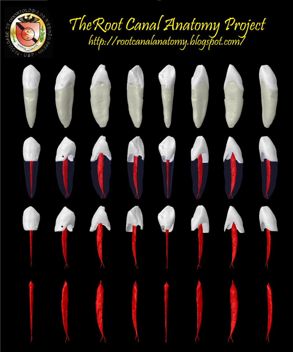
Narrow and  flat in the  labial-lingual  dimension, the mandibular incisors are the smallest human adult  teeth.  Visible radiographically  from  only one  plane,  they  often  appear more accessible than they really  are. The  narrow lingual crown offers a  limited area for  access. Smaller  fissure burs and no. 2  round burs  cause  less  mutilation of coronal  dentition. The access cavity  should be  ovoid,  with  attention given to a lingual approach. Frequently the mandibular  incisors have two  canals. One study  reported  that  41.4% of mandibular incisors studied had two separate  canals; of  these,  only  1.3% had two  separate foramina. The  clinician should  search for the  second  canal immediately upon  completing the access  cavity. Endodontic  failures in mandibular incisors usually  arise from  uncleaned canals, most commonly  toward the  lingual.  Access may be extended incisally when indicated to permit maximum  labiallingual  freedom.  Although labial  perforations are  common,  they may be avoided if the clinician  remembers  that it is nearly  impossible to perforate in a  lingual  direction because of  the bur shank's contacting the incisal edge. The ribbon-shaped canal is common enough to be considered normal and  demands special  attention in cleaning and shaping. Ribbon-shaped  canals in narrow  hourglass  cross-sectional anatomy invite  lateral perforation  by endodontic  files and Gates-Glidden  drills.  Minimal  flaring and  dowel space  preparation are indicated to  ensure  against ripping  through  proximal root walls. Apical curvatures and accessory canals are common in mandibular incisors (Burns RC, Buchanan LS. Tooth Morphology and Access Openings. Part One: The Art of Endodontics in Pathway of Pulp, 6th Ed. p. 152).


Keywords: micro-computed tomography, micro-ct, marco versiani, micro-computer tomography, high resolution x-ray tomography, dental anatomy, root canal anatomy