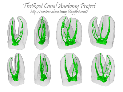
From 2011-2016, images and videos of "The Root Canal Anatomy Project" were developed at the Laboratory of Endodontics of Ribeirao Preto Dental School. From 2016, images were acquired in other educational institutions. They can be freely used for attributed noncommercial educational purposes by educators, scholars, student and clinicians. It means that all material used should include proper attribution and citation (http://rootcanalanatomy.blogspot.com). In such cases, this information should be linked to the image in a manner compatible with such instructional objectives. Unfortunately, because material shared on the RCAP has not been properly cited by several users, from November 2019 a watermark was added to the images and videos. Enjoy!

April 10, 2013

Roots Magazine 01.2013


Cover: 3-D micro-CT models of a mandibular molar showing  the changes of the original root-canal anatomy (green) after preparation with a multiple-file rotary system. Each color  represents preparation by one of  five  instruments. The  last  image  in the sequence  represents the  root canal after shaping  (red) superimposed on  the original canal  (green), demonstrating  that most of  the surface area was prepared using  the multiple-file system.

Click on the picture to enlarge

Thanks to Oemus (in the name of Magda Wojtkiewicz) for the invitation to write the Editorial of this issue of Roots magazine as well as to illustrate the cover. I hope you also enjoy it! 

Best regards,

Prof. Dr. Marco Versiani

March 16, 2013

Middle Mesial Canal: Maxillary First Molar


Click on the picture to enlarge
Source: Pathways of Pulp 10th Ed (p.190)


Middle Mesial Canal: Mandibular First Molar


Click on the picture to enlarge
Source: Pathway of Pulp 10th Ed. (p. 209)

In a study of 760 mandibular molars, Fabra found that 20 (2.6%) had three canals in the mesial root. In 13 of these (65%) the third canal joined the mesiobuccal canal in the apical third of the root and in 6 (30%) they converged with the mesiolingual canal, also in the apical third; the third canal ended as an independent canal in only 1 case. Many authors agree on the presence of three foramena in the mesial root but few report three independent canals, which presents itself as a rare anatomical variant. Walker cites 3 canals in the mesial root of the mandibular first molars as an infrequent ocurrence. Goel notes that the mesial root of permanent mandibular first molars presented two foramena in 60% of the specimens they examined, 6,7% had three and 3.3% even had four. In early studies by Hess, only 1 out of 55 teeth presented 2 distal roots and 5 canals did not appear in any of the teeth. However, many other authors confirm the hypothesis of a third canal in the mesial root of the permanent mandibular first molars According to Mortman, the third mesial canal is not an extra canal but rather the sequelae of instrumenting the isthmus between the mesiobuccal and mesiolingual canals. The isthmus is a narrow connection between two root canals that contains pulp tissue. In clinical practice, this isthmus is important in the conventional treatment of canals and in periapical surgery. In both cases it can lead to failure because it makes the canals very difficult to shape. An isthmus is located in between 54% and 89% of cases, most frequently between 4 mm and 6 mm from the apical foramen. The studies that examine series of cross-sections taken at different distances from the apex observe that isthmuses mainly occur at 3-5 mm from the apex. The prevalence and types of isthmus that can be identified in first molars by endoscopic examination during periapical surgery are as follows for the mesial root of the mandibular first molar: in a sample of 52 mesial roots, a canal with no isthmus was found in 3 cases (6%), two canals with no isthmus in 6 (11%) and two canals with isthmus in 43 (83%). Read more here. 


March 10, 2013

The Root Canal Anatomy Project: Two Years Online

Begun formally in 2011, The Root Canal Anatomy Project completed two years online on 5th of March. This blog was developed in the Laboratory of Endodontics of Ribeirao Preto Dental School - University of Sao Paulo - with noncommercial and educational purposes. During this period, the blog was visited by people from 163 different countries with more than 221,000 page views. The videos were watched more that 54,000 times and the material was downloaded more than 6,000 times. Considering that root canal anatomy is a very specific subject in the endodontic field, we believe that the blog is achieving its goals. Thank you for your support and stay tuned for updates.

Root Canal Anatomy Project
Statistics From March 2011 to March 2013


January 28, 2013

Single-File Technique Sample 1

Single-File Technique Series

The introduction of nickel-titanium (NiTi) rotary file systems has resulted in a markedly progress on the mechanical preparation of the root canal space. NiTi instruments offer many advantages over conventional stainless steel files such as flexibility and cutting efficiency. Furthermore, NiTi instruments maintain the original canal shape during preparation and have reduced tendency to transport the apical foramen. However, the current technology for mechanical preparation - rotary NiTi files - have failed in debriding oval-shaped canals, leaving untouched fins or recesses on the buccal and/or lingual extensions. This untouched recesses may harbor unaffected residual bacterial biofilms and serve as a potential cause of persistent infection and poor treatment outcome. Besides, as these techniques also require the use of numerous instruments to enlarge the canal to an adequate size and taper, they are relatively time and cost consuming.
The newly developed reciprocating NiTi files are made of a special NiTi alloy (M-Wire) and are claimed to be able to mechanically prepare the root canal space with only one instrument. These files area available in different sizes, indicated to be used according to the initial canal diameter. Initial reports using these instruments in extracted teeth have shown that they can debride the root canal space similarly to conventional multi-instrument rotary systems.
Several methodologies were developed to evaluate the shaping ability of NiTi systems, including simulated root canal models, serial sectioning technique, and radiographic comparison. These methodologies have been successfully used for many years; however, some inherent limitations encouraging the search for new methods able to produce improved results. The development of X-ray micro-computed tomography (µCT) has gained increasing significance in the study of dental tissues. µCT offers a noninvasive reproducible technique for three-dimensional assessment of the root canal system and it can be applied both quantitatively and qualitatively.
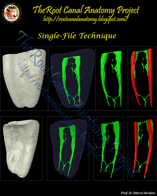
The purpose of this single-file technique series is to provide some material for educational purposes which, in summary, shows that is the root canal anatomy that dictates the result of the instrumentation procedure and not the type of instrument or system used. Besides, it calls the attention to the importance of the irrigation during root canal preparation.
  
Click here to download an editable Power Point file with all pictures and movies 

Mesial root of mandibular molar The 3D model in GREEN indicates the original root canal anatomy. The 3D model in RED indicates the root canal modified after preparation with a single-file system superimposed to the original canal.

Single-File Technique Sample 2


Mesial root of mandibular molar -  The 3D model in GREEN indicates the original root canal anatomy. The 3D model in RED indicates the root canal modified after preparation with a single-file system superimposed to the original canal.

Single-File Technique Sample 3


Mesial root of mandibular molar -  The 3D model in GREEN indicates the original root canal anatomy. The 3D model in RED indicates the root canal modified after preparation with a single-file system superimposed to the original canal.

Single-File Technique Sample 4


Mesial root of mandibular molar -  The 3D model in GREEN indicates the original root canal anatomy. The 3D model in RED indicates the root canal modified after preparation with a single-file system superimposed to the original canal.

Single-File Technique Sample 5


Mesial root of mandibular molar -  The 3D model in GREEN indicates the original root canal anatomy. The 3D model in RED indicates the root canal modified after preparation with a single-file system superimposed to the original canal.

Single-File Technique Sample 6


Mesial root of mandibular molar -  The 3D model in GREEN indicates the original root canal anatomy. The 3D model in RED indicates the root canal modified after preparation with a single-file system superimposed to the original canal.