From 2011-2016, images and videos of "The Root Canal Anatomy Project" were developed at the Laboratory of Endodontics of Ribeirao Preto Dental School. From 2016, images were acquired in other educational institutions. They can be freely used for attributed noncommercial educational purposes by educators, scholars, student and clinicians. It means that all material used should include proper attribution and citation (http://rootcanalanatomy.blogspot.com). In such cases, this information should be linked to the image in a manner compatible with such instructional objectives. Unfortunately, because material shared on the RCAP has not been properly cited by several users, from November 2019 a watermark was added to the images and videos. Enjoy!

March 6, 2014

Mandibular First Molar (with Isthmuses)


Mandibular First Molar (Vertucci Type IV)


Publication Eur Arch Paediatr Dent

Abstract
Aim 
This was to investigate the root canal morphology of primary molar teeth using micro-computed tomography.

Methods 
Primary maxillary (n=20) and mandibular (n=20) molars were scanned and analysed regarding the number, location, volume, area, structured model index (SMI), area, roundness, diameters, and length of canals, as well as the thickness of dentine in the apical third. Data were statistically compared by using paired-samplet test, independent sample t test, and one-way analysis of variance with significance level set as 5 %. 

Results 
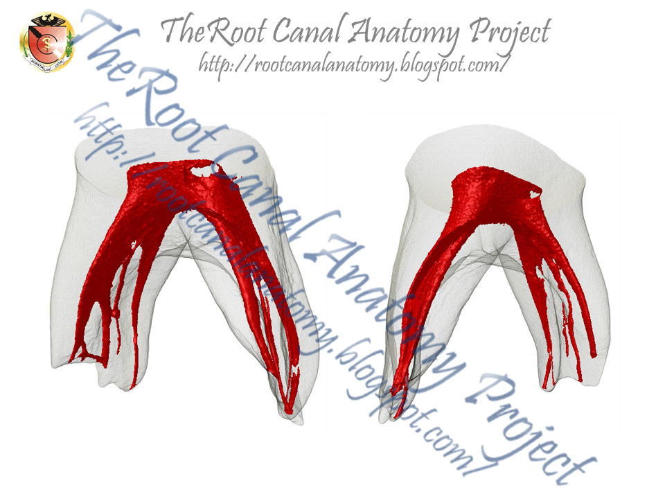
Overall, no statistical differences were found between the canals with respect to length, SMI, dentine thickness, area, roundness, and diameter (p<0.05). A double canal system was observed in the mesial and mesio-buccal roots of the mandibular and maxillary molars, respectively. The thickness in the internal aspect of the roots was lower than in the external aspect. Cross-sectional evaluation of the roots in the apical third showed flat-shaped canals in the mandibular molars and ribbon- and oval-shaped canals in the maxillary molars. Conclusions External and internal anatomy of the pri-mary first molars closely resemble the primary second molars. The reported data may help clinicians to obtain a
thorough understanding of the morphological variations of root canals in primary molars to overcome problems rela-ted to shaping and cleaning procedures, allowing appro-priate management strategies for root canal treatment.

Conclusions 
External and internal anatomy of the pri-mary first molars closely resemble the primary second molars. The reported data may help clinicians to obtain a thorough understanding of the morphological variations of root canals in primary molars to overcome problems related to shaping and cleaning procedures, allowing appropriate management strategies for root canal treatment.


December 10, 2013

Roots Magazine 04.2013

Frontal and lateral views of a 3-D reconstruction of a maxillary first premolar showing a three-rooted canal system. This micro-CT image was developed as part of the Root Canal Anatomy Project  in the Laboratory of Endodontics of the University of Sao Paulo in Ribeirao Preto, Brazil by Prof. Marco Versiani, Prof. Jesus Pécora & Prof. Manoel Sousa-Neto.


October 21, 2013

Publication JOE


Abstract 
Introduction
This study aimed to describe the anatomy of mandibular central and lateral incisors using micro–computed tomographic imaging.

Methods
One hundred mandibular incisors were scanned in a micro–computed tomographic device using an isotropic resolution of 22.9 μm. The anatomy of each tooth (length of the roots, presence and location of accessory canals and apical deltas, and number of canals) as well as the 2- and 3-dimensional morphologic aspects of the canal (area, roundness, diameter, volume, surface area, and structure model index) were evaluated. Data were statistically compared using the Student t test (alpha = 0.05).

Results
The mean lengths of the mandibular central and lateral incisors were 20.71 and 21.56 mm, respectively. Most of the central (60%) and lateral (74%) incisors had no accessory canals. An apical delta was observed in only 1 specimen. The cross-section analysis of the apical third showed the presence of 1, 2, or 3 canal orifices. No statistical difference was observed in the comparison of the 2- and 3-dimensional morphologic parameters between central and lateral incisors (P < .05). The qualitative analyses of the 3-dimensional models of the root canal systems of the central and lateral incisor teeth confirm that the most prevalent configurations were Vertucci's types I (50% and 62%, respectively) and III (28%).

Conclusions
Overall, mandibular central and lateral incisors were similar in terms of the 2- and 3-dimensional analyzed parameters. Vertucci's types I and III were the most prevalent canal configurations of the mandibular incisors; however, 8 new types have also been described.

Publication JOE


Abstract 
Introduction
The accumulation of debris occurs after root canal preparation procedures specifically in fins, isthmus, irregularities, and ramifications. The aim of this study was to present a step-by-step description of a new method used to longitudinally identify, measure, and 3-dimensionally map the accumulation of hard-tissue debris inside the root canal after biomechanical preparation using free software for image processing and analysis.

Methods
Three mandibular molars presenting the mesial root with a large isthmus width and a type II Vertucci's canal configuration were selected and scanned. The specimens were assigned to 1 of 3 experimental approaches: (1) 5.25% sodium hypochlorite + 17% EDTA, (2) bidistilled water, and (3) no irrigation. After root canal preparation, high-resolution scans of the teeth were accomplished, and free software packages were used to register and quantify the amount of accumulated hard-tissue debris in either canal space or isthmus areas.

Results
Canal preparation without irrigation resulted in 34.6% of its volume filled with hard-tissue debris, whereas the use of bidistilled water or NaOCl followed by EDTA showed a reduction in the percentage volume of debris to 16% and 11.3%, respectively. The closer the distance to the isthmus area was the larger the amount of accumulated debris regardless of the irrigating protocol used.

Conclusions
Through the present method, it was possible to calculate the volume of hard-tissue debris in the isthmuses and in the root canal space. Free-software packages used for image reconstruction, registering, and analysis have shown to be promising for end-user application.

September 28, 2013

Why does mandibular incisor fail?

Usually, teeth with single roots present single canals as in mandibular and maxillary anterior teeth. However, particular tooth types, such as mandibular premolars and incisors, are recognized as exhibiting a distinct range of variations in the morphology of their root canal system. In mandibular incisors, often a dentinal bridge is present in the pulp chamber dividing the root into two canals. The two canals usually join and exit through a single apical foramen, but they may persist as two separate canals. On occasion one canal branches into two canals, which subsequently rejoin into a single canal before reaching the apex. The incidence of two canals in mandibular incisors has been reported to be as low as 0.3% and as high as 45.3%. The wide range of variation reported in the literature regarding the prevalence of a second canal in mandibular incisors has been mostly related to methodological and racial differences.
(Very soon on the Journal of Endodontics)