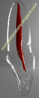
In the last decade, micro-CT has gained increasing popularity in endodontics. This noninvasive, nondestructive, high-resolution technology allows the three-dimensional study of the root canal system and can be used to understand its influence on the different treatment/retreatment procedures, by reconstructing digital cross sections of the teeth, which can be stacked to create 3D volumes. These volumes can be used to generate computerized images of specimens that can be manipulated, or measured, to reveal both internal and external morphologies. Nowadays, micro-CT technology is considered the most important and accurate research tool to the study of root canal anatomy. Images below were acquired in a micro-CT device (so, they are based on real teeth) and processed with dedicated modelling 3D software for the book ROOT CANAL ANATOMY IN PERMANENT DENTITION.
Additional information regarding the use micro-CT technology in endodontics can be found in this book CHAPTER.


No comments:
Post a Comment