From 2011-2016, images and videos of "The Root Canal Anatomy Project" were developed at the Laboratory of Endodontics of Ribeirao Preto Dental School. From 2016, images were acquired in other educational institutions. They can be freely used for attributed noncommercial educational purposes by educators, scholars, student and clinicians. It means that all material used should include proper attribution and citation (http://rootcanalanatomy.blogspot.com). In such cases, this information should be linked to the image in a manner compatible with such instructional objectives. Unfortunately, because material shared on the RCAP has not been properly cited by several users, from November 2019 a watermark was added to the images and videos. Enjoy!
Showing posts with label Mandibular Canine. Show all posts

May 1, 2024

Worldwide studies: Mandibular Canine

 Worldwide Anatomic Characteristics of the Mandibular Canine-A Multicenter Cross-Sectional Study with Meta-Analysis

Jorge N R Martins; Worldwide Anatomy Research Group; Marco A Versiani

Link to the original publication

Click here to read the paper


January 13, 2013

Mandibular Canine


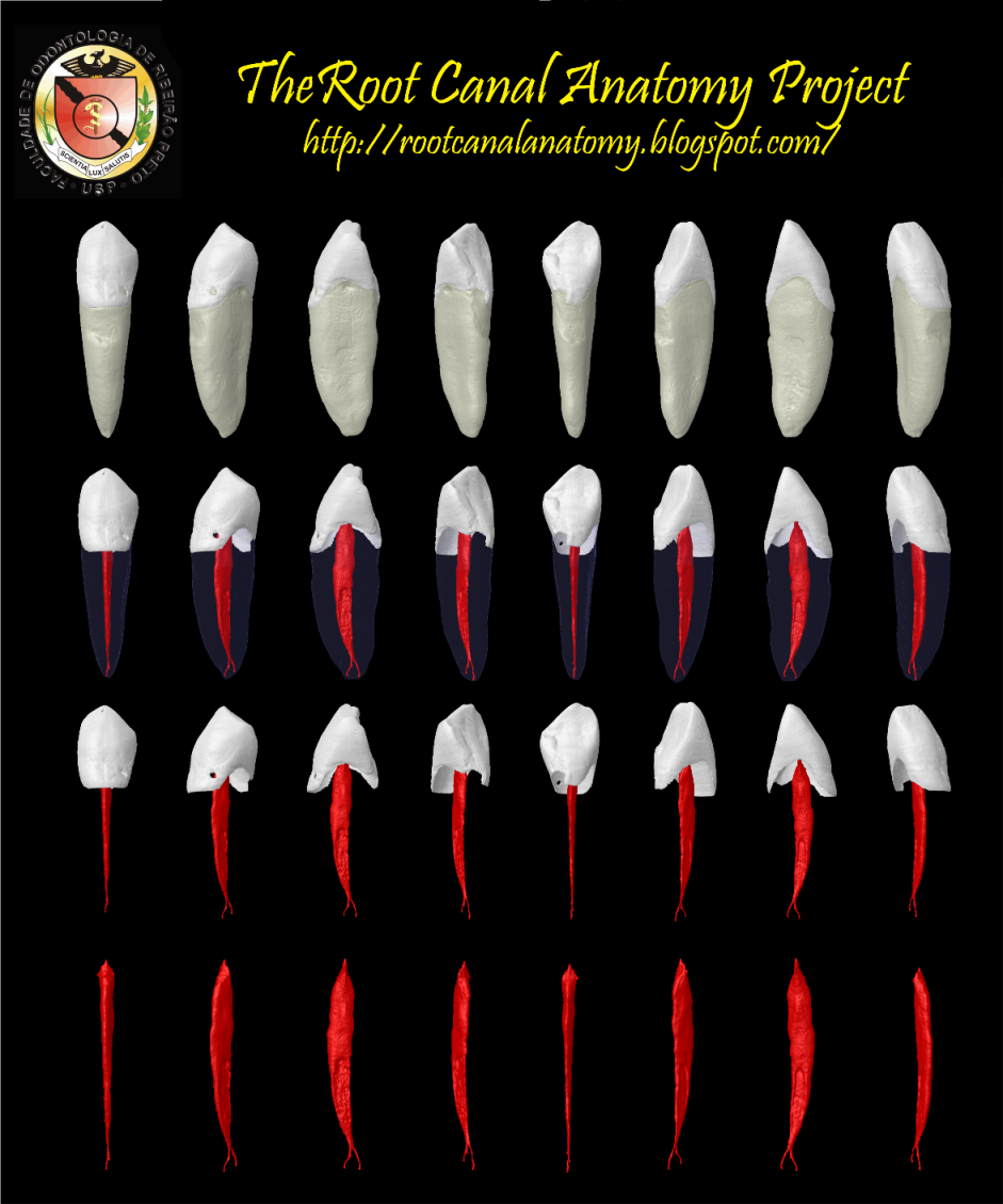
The article entitled "Microcomputed tomography study of the root canal morphology of single-rooted mandibular canine" was published on the International Endodontic Journal. By now, I am going to provide you with some pictures and a movie of this group of teeth. 
P.S. The original article contains different pictures from the below.


May 12, 2012

Two-Rooted Mandibular Canine

Although mandibular canine usually have one root canal, the occurrence of two roots and two distinct
canals has often been reported (Ouellet 1995, Sharma et al. 1998, D’Arcangelo et al. 2001, Victorino et al. 2009). Most reports refer to two-rooted mandibular canine in case reports (D’Arcangelo et al. 2001, Victorino et al. 2009), whilst data from ex vivo studies report this anatomic variation to occur in 1.7% (Pecora et al. 1993) to 5% (Ouellet 1995). READ MORE.


This video contains the three-dimensional resolution as well as the axial sections of the tooth

Keywords: micro-computed tomography, micro-ct, marco versiani, micro-computer tomography, high resolution x-ray tomography, dental anatomy, root canal anatomy

April 6, 2011

Mandibular canine

Sturdy and  considerably wider mesial-distally than the incisors, the  mandibular canines...read more. 


Keywords: micro-computed tomography, micro-ct, marco versiani, micro-computer tomography, high resolution x-ray tomography, dental anatomy, root canal anatomy

March 4, 2011

Mandibular Canine

Sturdy and  considerably wider mesial-distally than the incisors, the  mandibular canines  seldom present  endodontic problems. The  unusual occurrence of  two roots can  create  difficulty, but this is  rare. The access  cavity is ovoid and  may be  extended  incisally for labial-lingual  accessibility. The canal is  somewhat  ovoid at the  cervical,  becoming  round at midroot.  Directional  instrumentation is necessary  to debride the  canal walls  completely. If there are two  roots, one is  always easier to  instrument. The other must be opened and  funneled in concert with the first to prevent packing of dentin debris and loss of access. Precurving of instruments at  initial  access  will  enable the clinician to trace down the buccal or  lingual root  wall until the tip engages the  orifice. When the  difficult canal is located, every  effort  should be made to shape and  funnel the opening to  maintain  continued  access (Burns RC, Buchanan LS. Tooth Morphology and Access Openings. Part One: The Art of Endodontics in Pathway of Pulp, 6th Ed. p. 154).


Keywords: micro-computed tomography, micro-ct, marco versiani, micro-computer tomography, high resolution x-ray tomography, dental anatomy, root canal anatomy