From 2011-2016, images and videos of "The Root Canal Anatomy Project" were developed at the Laboratory of Endodontics of Ribeirao Preto Dental School. From 2016, images were acquired in other educational institutions. They can be freely used for attributed noncommercial educational purposes by educators, scholars, student and clinicians. It means that all material used should include proper attribution and citation (http://rootcanalanatomy.blogspot.com). In such cases, this information should be linked to the image in a manner compatible with such instructional objectives. Unfortunately, because material shared on the RCAP has not been properly cited by several users, from November 2019 a watermark was added to the images and videos. Enjoy!
Showing posts with label Mandibular First Molar. Show all posts

March 3, 2023

Mandibular First Molar

Root Canal Anatomy Project 

Merging Science & Art

Realistic 3D model obtained by micro-CT technology and characterized by using advanced computer design techniques. Watch in HD!

October 3, 2020

Root Canal Anatomy: 3D animations

Root Canal Anatomy Project

Merging Art & Science

Flying over the root canal system of a mandibular first molar


Sometimes, anatomy makes me crazy


If the supervillain Thanos from Marvel decided that root canal should die


The anatomy and the heat-treated instrument are based on real objects (microCT-based model).
But - in clinics - would it be able to do that?

Root canal system of a maxillary first molar


C-shaped canal in a mandibular second molar

December 1, 2019

Middle Mesial Canal: Rotary Preparation


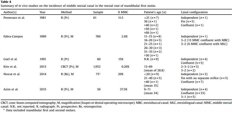
The mesial root of mandibular molars commonly presents 2 main root canals [(mesiobuccal (MB) and mesiolingual (ML)], but the presence of an extra canal in this root, the so-called middle mesial (MM) canal, has been also reported in a percentage frequency ranging from 0.26% to 46.15% . Considering that MM canal lies within a thin developmental groove between the orifices of the MB and ML canals, troughing this groove under high magnification has been suggested in order to identify its presence. However, in a detailed morphological description of the mesial root of mandibular molars presenting MM canal, authors stated that the presence of a thin dentine thickness toward the furcation side of the MM canal at the orifice level (0.80–2.20 mm) would  increase the risk of root perforation after preparation with large-tapered instruments.

For more information click HERE or HERE or HERE


December 12, 2015

Publication: Comparative accuracy of the Clearing Technique, CBCT and Micro-CT

COMPARATIVE ACCURACY OF THE CLEARING TECHNIQUE, CBCT, AND MICRO-CT METHODS IN STUDYING MESIAL ROOT CANAL CONFIGURATION OF MANDIBULAR FIRST MOLARS


R Ordinola-Zapata, C M. Bramante, M A. Versiani, I B Moldauer, G Topham, 
J L. Gutmann, A Nuñez, M A Hungaro Duarte and F Abella

Despite the considerable number of studies published on the internal anatomy of posterior teeth, very little information exists regarding the accuracy of clearing, CBCT, and micro-CT methods to diagnose the morphology of the root canal anatomy. Therefore, the aim of this study was to compare the accuracy of the clearing technique and CBCT scanning in the assessment of the mesial root canal configuration of mandibular first molars, using micro-CT imaging system as a reference standard. 


November 8, 2015

Publication: Middle Mesial Canal

Middle mesial canals in mandibular first molars: 
a micro-CT study in different populations

Marco Aurélio Versiani, Ronald Ordinola-Zapata, Ali KeleÅŸ, Hatice Alcin, 
Clóvis Monteiro Bramante, Jesus Djalma Pécora, Manoel Damião Sousa-Neto

Objective
To describe the morphological aspects of middle mesial canals (MMC) in mandibular first molars using micro-CT.

Design
Mandibular first molars collected from the Brazilian (n=136) and Turkish (n=122) populations were scanned (voxel size: 9.9 μm) and mesial roots with MMC (n=48) evaluated regarding several morphological aspects. The incidence of MMC in each population was statistically compared using Chi-square test (α=0.05).

Results
Overall, the incidence of MMC was 18.6% (48 out of 258 molars) and was significantly higher in the Brazilian (n=30; 22.1%) than in the Turkish (n=18; 14.8%) population (p<0.05). In both populations, confluent configuration of the MMC was the most frequent anatomy. Most of the specimens with MMC had 3 independent orifices (n=26; 54.2%) and 3 apical foramina (n=21; 43.8%). The mean minor diameter of the MMC orifice (0.16 mm) was 3 times less than the other orifices (∼0.50 mm). In mesial roots with independent configuration (n=3; 6.3%), the mean volumes (mm3) of the MMC, mesiobuccal (MBC) and mesiolingual (MLC) canals were 0.20±0.10, 0.75±0.28, and 0.88±0.19, respectively. In the specimens with canal confluence (n=26; 54.2%), MMC merged to the MBC (n=8; 16.7%), MLC (n=4; 8.3%), or to both MBC and MLC (n=14; 29.2%). Double mesial canal was observed in only 1 specimen. MMC with an independent foramen was observed mostly in Brazilian specimens.

Conclusions
Incidence of MMC was higher in the Brazilian molars. Confluent configuration was the most revalent anatomic variation, while independent and fin configurations, as well as, double MMC, were found only in a few specimens.

Keywords
mandibular molar; middle mesial canal; micro-CT; root canal anatomy

Click HERE or HERE to read and download the article in full


July 11, 2015

Middle Mesial Canal - Mandibular Molar


Middle Mesial Canal - Mandibular Molar


Middle Mesial Canal - Mandibular Molar


Middle Mesial Canal - Mandibular Molar


Middle Mesial Canal - Mandibular Molar


February 1, 2015

Root Canal Anatomy X Rotary Preparation

The influence of the anatomical configuration of the root canal on the root canal preparation

Type IV (Vertucci's Configuration System)

Type V (Vertucci's Configuration System)

Watch this video in HD!

November 16, 2014

Root Canal Preparation: Mandibular Molar

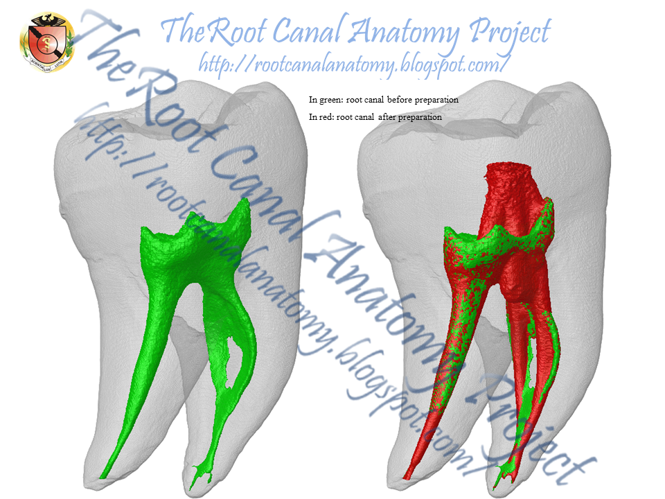
Root Canal Preparation of a Mandibular Molar 
Using a Multiple-File Rotary NiTi System

Thanks to Dr. Ali Keles from Turkey for the scanning and canal preparation procedures
In green: root canal before preparation
In red: root canal after preparation superimposed to the original canal


August 10, 2014

Conservative Endodontic Access - Mandibular Molar

Conservative Endodontic Access - Mandibular Molar

The principles of traditional endodontic cavities were established many decades ago and have remained unchanged over time. The principles of traditional endodontic cavities were established many decades ago and have remained unchanged over time. The  emerging concept of conservative endodontic access prioritizes the removal of restorative materials before tooth structure, of enamel before dentin and of occlusal tooth structure before cervical dentin. It disregards the traditional requirements of a straight-line access and complete unroofing of the pulp chamber and it emphasizes the importance of preservation of the crucial pericervical dentin (located 4 mm above and below the crestal bone) to the greatest extent possible.