From 2011-2016, images and videos of "The Root Canal Anatomy Project" were developed at the Laboratory of Endodontics of Ribeirao Preto Dental School. From 2016, images were acquired in other educational institutions. They can be freely used for attributed noncommercial educational purposes by educators, scholars, student and clinicians. It means that all material used should include proper attribution and citation (http://rootcanalanatomy.blogspot.com). In such cases, this information should be linked to the image in a manner compatible with such instructional objectives. Unfortunately, because material shared on the RCAP has not been properly cited by several users, from November 2019 a watermark was added to the images and videos. Enjoy!

November 8, 2020

Radix Entomolarix (Mandibular Molar)


Root Canal Anatomy Project Merging Art & Science
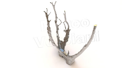
Three-dimensional microCT-based root canal model of a mandibular second molar with radix entomolaris using advanced computacional design tools

October 26, 2020

Maxillary Lateral Incisor


Root Canal Anatomy Project
Merging Science & Art

Realistic 3D model obtained by micro-CT technology and characterized by using advanced computer design techniques.

October 3, 2020

Worldwide expertise in X-Ray Micro-CT


Realistic 3D Anatomies

Root Canal Anatomy Project
Merging Art & Science

Realistic 3D models obtained with micro-CT technology and textured using advanced computacional graphic methods.


Root Canal Anatomy: 3D animations

Root Canal Anatomy Project

Merging Art & Science

Flying over the root canal system of a mandibular first molar


Sometimes, anatomy makes me crazy


If the supervillain Thanos from Marvel decided that root canal should die


The anatomy and the heat-treated instrument are based on real objects (microCT-based model).
But - in clinics - would it be able to do that?

Root canal system of a maxillary first molar


C-shaped canal in a mandibular second molar

May 17, 2020

May 16, 2020