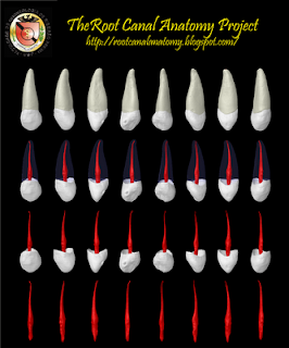
The longest tooth in the dental arch, the canine has a formidable shape designed to withstand heavy occlusal stress. Its long, thickly enameled crown sustains heavy incisal wear but often displays deep cervical erosion with aging. The access cavity corresponds to the lingual crown shape and is ovoid. To achieve straight-line access, one often must extend the cavity incisally, but not so far as to weaken the heavily functioning cusp. Initial access is made slightly below midcrown on the palatal side. If the pulp chamber is located deeper, a no. 4 or 6 long-shanked round bur may be required. The sweeping-out motion of this bur will reveal an ovoid pulp chamber. The chamber remains ovoid as it continues apically through the cervical region and below. Attention must be given to directional tiling so this ovoid chamber will be thoroughly cleaned. The radicular canal is reasonably straight and quite long. Most canines require instruments that are 25 mm or longer. The apex will often curve—in any direction—in the last 2 or 3 mm. Canine morphology seldom varies radically, and lateral and accessory canals occur less frequently than in the maxillary incisors. This buccal bone over the eminence often disintegrates, and fenestration is a common finding. The apical foramen is usually close to the anatomic apex but may be laterally positioned, especially when apical curvature is present (Burns RC, Buchanan LS. Tooth Morphology and Access Openings. Part One: The Art of Endodontics in Pathway of Pulp, 6th Ed. p. 140).
Keywords: micro-computed tomography, micro-ct, marco versiani, micro-computer tomography, high resolution x-ray tomography, dental anatomy, root canal anatomy


No comments:
Post a Comment