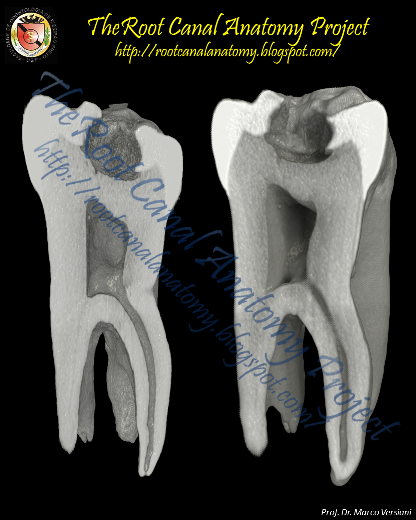
From 2011-2016, images and videos of "The Root Canal Anatomy Project" were developed at the Laboratory of Endodontics of Ribeirao Preto Dental School. From 2016, images were acquired in other educational institutions. They can be freely used for attributed noncommercial educational purposes by educators, scholars, student and clinicians. It means that all material used should include proper attribution and citation (http://rootcanalanatomy.blogspot.com). In such cases, this information should be linked to the image in a manner compatible with such instructional objectives. Unfortunately, because material shared on the RCAP has not been properly cited by several users, from November 2019 a watermark was added to the images and videos. Enjoy!

September 23, 2012

Impressions from Warwick Medical School

Supervisors Training Workshop (Dissertation and Professional Project) at the Warwick Medical School (University of Warwick) in 11th September 2012. A personal thanks to Dr. Liviu Steier for the invitation and Dr. Edward Lynch for the warm reception. Amazing day, surrounded by amazing people. Besides, I had the virtual company of some nice guys as Antonis Chaniotis, Freddy Belliard, Roberto Cristescu, and others. Living and learning...


September 20, 2012

APCD Journal Interview


França S. Avanços no desenvolvimento de materiais e técnicas aperfeiçoam o controle da infecção endodôntica. Rev Assoc Paul Cir Dent 2012;66(3):166-71 (In Portuguese Only)
[Development of techniques and materials to improve the control of the endodontic infection]
Prof. Marco Versiani, Prof. Gustavo de Deus, Prof. Ghassan Yared, Prof. Giulio Gavini


August 11, 2012

Four-Rooted Maxillary Second Molars

None of the present pictures were previously published in the
cited article below from Journal of Endodontics


The video from all these teeth are available as a
supplemental material on JOE's website


August 5, 2012

Maxillary First Molar: Five Canals

3D Reconstruction of a Maxillary First Molar with Five Root Canals
Micro-CT Models: Carabelli's Tubercle in the MB cusp (read more)
 Micro-CT Models: Cervical Enamel Projection (read more)
Micro-CT Models: Overview of the Pulp Chamber (Canal Orifices)
 Micro-CT Models: Canal Orifices
Micro-CT Model: Palatal Canal

June 17, 2012

Impressions from VDW (Munich)

From left to right: Mr. Walter Loaiza (VDW Germany), Prof. Dr. Gianluca Plotino, Prof. Dr. Key Fabiano Souza Pereira, Prof. Dr. Eudes Gondim, Dra. Lilia Macedo França, Prof. Dr. Erick Miranda Souza, Prof. Dr. Flávio Rodrigues Ferreira Alves, Mr. Salomon Matto (VDW Brazil), Prof. Dr. Gustavo de Deus, Prof. Dr. Marco Versiani, Prof. Dr. José Mauricio Paradella de Camargo
 VDW Training Room
  VDW Training Room
VDW Training Room
  VDW Training Room
 Dr. Gustavo de Deus, Dr. Erick Miranda Souza, Dr. Gianluca Plotino, Dr. Marco Versiani
 The Brazilian Team with Dr. Gianluca Plotino (Center)
 
 Dr. Gustavo de Deus, Dr. Erick Miranda Souza, Dr. Marco Versiani
  Dr. Gustavo de Deus, Dr. Erick Miranda Souza, Dr. Marco Versiani
Dr. Erick Miranda Souza, Dr. Marco Versiani
A special thanks to VDW team: Mr. Walter Loaiza, Mrs. Lydia Bonenberger, Mrs. Pamela Marklew, Mr. Markus Borgschulte, Mrs. Corinna Ludwig, Mrs. Karin Duhnke, Mrs. Iris Köster, Mrs. Carolin Höschen

June 2, 2012

Root Canal Preparation: Gates-Glidden + K-Files

Root Canal Preparation Protocol (Crown-Down Technique):

Mesiobuccal and Mesiolingual Canals 
- Cervical Third (Gates-Glidden burs #3 - #1)
- Middle and Apical Thirds (Stainless Steel K-Files - Apical Preparation: K-File: #30)

Distal Canal
- Cervical Third (Gates-Glidden burs #2 - #4)
- Middle and Apical Thirds (Stainless Steel K-Files - Apical Preparation: K-File: #40)

Click on the images to enlarge

3D Models of Root Canal Before Preparation (Green)
3D Models of Root Canal After Preparation (Red)
3D Models of Root Canal Before and After Preparation
Registration Process

Five Transaxial Views Of The Root
Superimposition of the Canal Before and After Preparation
Superimposition of the 3D Models of the Root Canal
Before (Green) and After (Red) Canal Preparation

Cervical Preparation With Gates-Glidden Burs
Before (Black) and After (Red) Canal Preparation

Axial Cross-Sections of a Mandibular Molar
Root Canal Preparation (Crown-Down Technique)
[Gates-Glidden Burs + Stainless Steel K-Files]