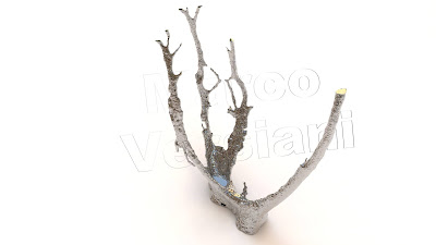
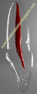
3D models of root canals textured with computer graphics to resemble pulp tissue
June 29, 2024
May 1, 2024
The Palato-Mesiobuccal Canal
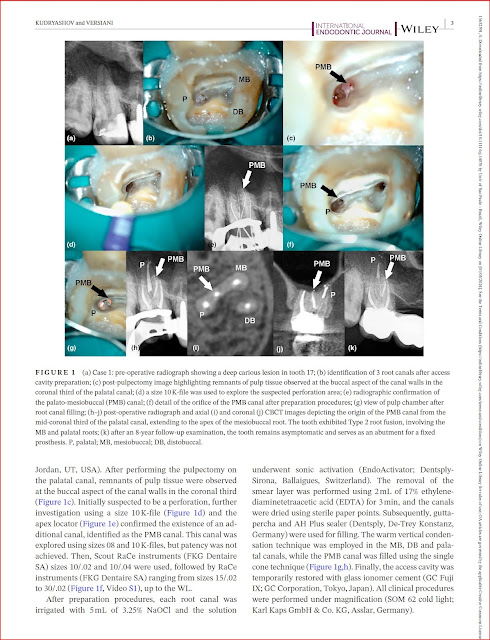
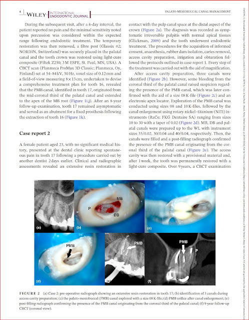
Identification and Characterization of a Previously Undiscovered Anatomical Structure in Maxillary Second Molars: The Palato-Mesiobuccal Canal
Marco A Versiani, Tamer Taşdemir, Ali Keleş
Link to the original publication
Anatomical Complexities of the Root Canal System
Anatomical complexities affecting root canal preparation: a narrative review
M A Versiani JNR Martins R Ordinola-Zapata
Link to the original publication
Pulp Calcification
Micro‑CT assessment of radicular pulp calcifcations in extracted maxillary frst molar teeth
Ali Keleş Cangül Keskin, Marco Versiani
Link to the original publication
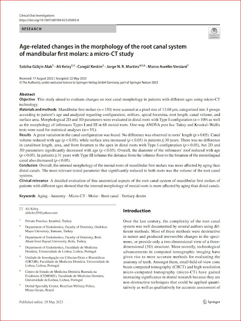
Age‑related changes in the morphology of the root canal system of mandibular frst molars: a micro‑CT study
Sabiha Gülçin Alak, Ali Keleş, Cangül Keskin, Jorge N. R. Martins, Marco Versiani,
Link to the original publication
March 3, 2023
Mandibular First Molar
Root Canal Anatomy Project
Merging Science & Art
Realistic 3D model obtained by micro-CT technology and characterized by using advanced computer design techniques. Watch in HD!
March 2, 2023
Double Maxillary Molar
August 20, 2022
Maxillary First Molar - Deep Split
August 19, 2022
Maxillary First Molar - Type II Configuration
The most common root canal system configuration of a maxillary first molar: 1 palatal, 1 distobuccal and 2 mesiobuccal (MB1 and MB2 canals with Vertucci's Type II configuration root canals. Photorealistic texture of a 3D model acquired using micro-CT technology and performed with advanced computational tools.
November 8, 2020
Radix Entomolarix (Mandibular Molar)
October 30, 2020
Mandibular Canine with Apical Ramification - Dr Marco Versiani
October 26, 2020
Maxillary Lateral Incisor
October 3, 2020
Root Canal Anatomy: 3D animations
Root Canal Anatomy Project
Merging Art & Science
Flying over the root canal system of a mandibular first molar


