3D models of root canals textured with computer graphics to resemble pulp tissue

Showing posts with label Maxillary First Premolar. Show all posts
Showing posts with label Maxillary First Premolar. Show all posts
June 29, 2024
May 1, 2024
Worldwide Studies: Maxillary Premolars
Worldwide Assessment of the Root and Root Canal Characteristics of Maxillary Premolars - A Multi-center Cone-beam Computed Tomography Cross-sectional Study With Meta-analysis
Jorge N R Martins; Worldwide Anatomy Research Group; Marco A Versiani
Link to the original publication


August 24, 2022
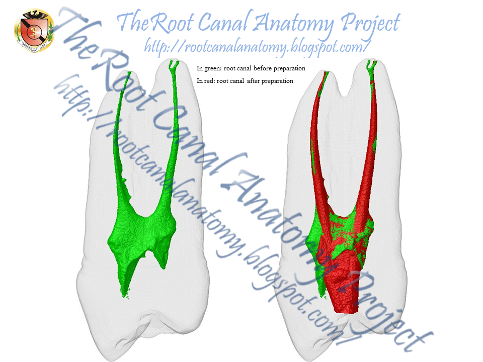
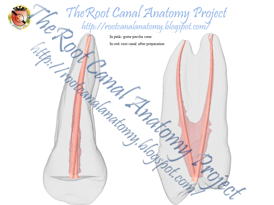
Three-rooted maxillary premolar
A new publication in the IEJ about root canal preparation of three-rooted maxillary premolars
Click here to read the article in full


November 24, 2019


October 30, 2015
Three-Rooted Maxillary Premolar
The following outstanding clinical case was performed by
Dr. Konstantinos Kalogeropoulos
and posted in the Facebook page 'Athens Endo'

Below, the anatomy of a similar three-rooted maxillary premolar is detailed


July 30, 2014
Conservative Endodontic Access - Maxillary Premolar
Conservative Endodontic Access - Maxillary Premolar
The principles of traditional endodontic cavities were established many decades ago and have remained unchanged over time. The principles of traditional endodontic cavities were established many decades ago and have remained unchanged over time. The emerging concept of conservative endodontic access prioritizes the removal of restorative materials before tooth structure, of enamel before dentin and of occlusal tooth structure before cervical dentin. It disregards the traditional requirements of a straight-line access and complete unroofing of the pulp chamber and it emphasizes the importance of preservation of the crucial pericervical dentin (located 4 mm above and below the crestal bone) to the greatest extent possible.
(for more information: Rajesh Krishan's Thesis from the University of Toronto)


May 24, 2014


August 20, 2013


October 1, 2011
Maxillary First Premolar
Most commonly birooted, the maxillary first premolar is a transitional tooth between incisor and molar. Read more.


Keywords: micro-computed tomography, micro-ct, marco versiani, micro-computer tomography, high resolution x-ray tomography, dental anatomy, root canal anatomy
March 4, 2011
Maxillary First Premolar
Most commonly birooted, the maxillary first premolar is a transitional tooth between incisor and molar. Loss of the posterior molars subjects the premolars to heavy occlusal loads. Removable appliances increase torque on these frequently clasped teeth, and the additional forces, in concert with deep carious lesions, can induce heavy calcification of the pulp chambers. Early posterior tooth loss often causes rotation, which can complicate the locating of pulp chambers. The canal orifices lie below and slightly central to the cusp tips. The initial opening is in the central fossa and is ovoid in the buccal-palatal dimension. When one orifice has been located, the clinician should look carefully for a developmental groove leading to the opening of another canal. The angulation of the roots may be determined by positioning of the endodontic explorer. Radiographic division of the roots on a routine periapical film often indicates tooth rotation. Divergent roots require less occlusal access extension. Conversely, parallel roots may require removal of tooth structure toward the cusp tips. All caries and leaking restorations must be removed and a suitable temporary restoration placed. Radicular irregularities consist of fused roots with separate canals, fused roots with interconnections or "webbing," fused roots with a common apical foramen, and the unusual but always to be considered three-rooted tooth. In the last situation the buccal orifices are not clearly visible with a mouth mirror. Directional positioning of the endodontic explorer or a small file will identify the anatomy. Cams and Skidmore reported that the incidence of maxillary first premolars with three roots, three canals, and three foramina was 6% of the cases studied. The root is considerably shorter than in the canine, and distal curvature is not uncommon. The apical foramen is usually close to the anatomic apex. Root lengths, if the cusps are intact and used as reference points, are usually the same. The apical portion of the roots often tapers rapidly, ending in extremely narrow and curved root tips. The prevalence of mesial-distal vertical crown or root fracture of the first premolar requires that the clinician remove all restorations at the inception of endodontic therapy and carefully inspect the coronal anatomy with a fiberoptic light. After endodontic treatment, full occlusal coverage is mandatory to ensure against vertical or crown or root fracture (Burns RC, Buchanan LS. Tooth Morphology and Access Openings. Part One: The Art of Endodontics in Pathway of Pulp, 6th Ed. p. 142).

Keywords: micro-computed tomography, micro-ct, marco versiani, micro-computer tomography, high resolution x-ray tomography, dental anatomy, root canal anatomy
Subscribe to:
Posts (Atom)