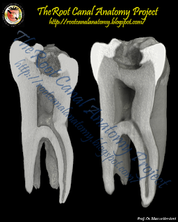
From 2011-2016, images and videos of "The Root Canal Anatomy Project" were developed at the Laboratory of Endodontics of Ribeirao Preto Dental School. From 2016, images were acquired in other educational institutions. They can be freely used for attributed noncommercial educational purposes by educators, scholars, student and clinicians. It means that all material used should include proper attribution and citation (http://rootcanalanatomy.blogspot.com). In such cases, this information should be linked to the image in a manner compatible with such instructional objectives. Unfortunately, because material shared on the RCAP has not been properly cited by several users, from November 2019 a watermark was added to the images and videos. Enjoy!
Showing posts with label Maxillary Second Molar. Show all posts

May 1, 2024

The Palato-Mesiobuccal Canal

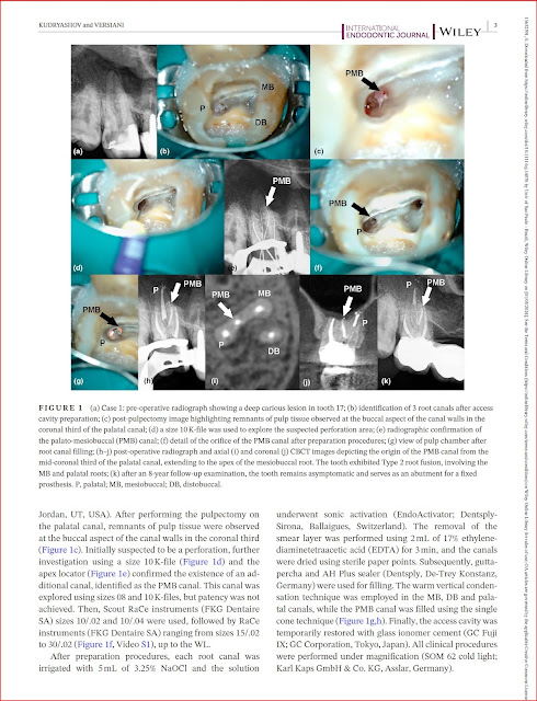
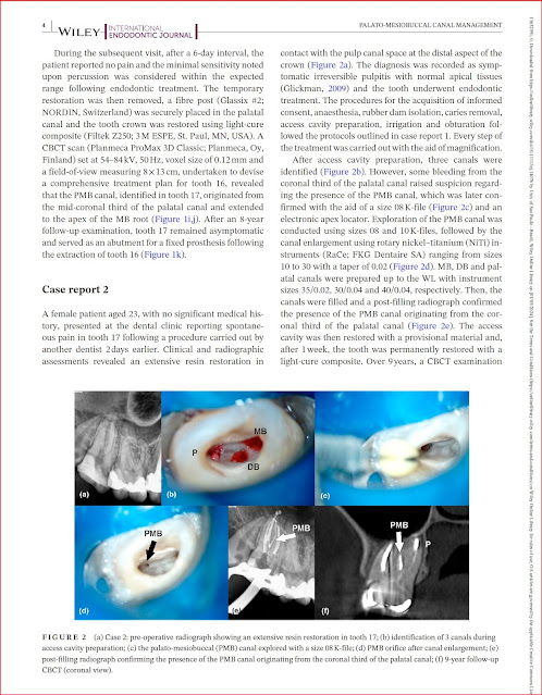
The palato-mesiobuccal canal, a new anatomical variation of maxillary second molars with fused roots, was described for the first time in the literature by our group. In the following links you can find additional information about its morphology and clinical management: 


 Identification and Characterization of a Previously Undiscovered Anatomical Structure in Maxillary Second Molars: The Palato-Mesiobuccal Canal

Marco A Versiani, Tamer Taşdemir, Ali Keleş

Link to the original publication

Click here to read the paper


Clinical strategies for successful palato-mesiobuccal canal management-Report of 2 cases

Dmitry Kudryashov, Marco A Versiani


December 12, 2019

MB2 canal: Systematic Review and Meta-Analysis


You can read the full text by clicking HERE, HERE or HERE

Summary

Prevalence studies using CBCT technology on MB2 canal were searched between May and September 2019. 83 studies were submitted to full text analysis and scientific merit evaluation by 2 evaluators and 26 studies were pooled into a meta-analysis. The included studies reported data of 23,926 maxillary molars (15,285 maxillary first molars and 8,641 maxillary second molars) from at least 12,456 patients, comprising 5,541 males and 6,915 females (2 studies did not report the number of patients). The average age of the patients was 40.9 years and was calculated based on 20 studies that reported this information. The included studies were published in English (n=24), Chinese (n=1) and Portuguese (n=1) and represented data from 24 countries.
Overall prevalence of MB2 canal

In the present study, prevalence of MB2 canal in maxillary first molars ranged from 96.7% (Belgium sub-population) to 30.9% (Chinese sub-population) while in second molars, the highest and lowest prevalence were reported in the Brazilian (83.2%) and Chinese (13.4%) sub-populations. Overall, mean prevalence of MB2 was higher in maxillary first molars (69.6%) than in second molars (39.0%). The presence of MB2 canal in maxillary first molars were addressed in 22 studies (41 population groups) with a high heterogeneity values for both maxillary first and second molars.

MB2 canal and gender

Influence of gender on the prevalence of MB2 canal in maxillary first molars was compared in 16 studies (35 population groups). Statistical comparison of untransformed proportions of MB2 for males (71.9%; 66.5%-77.4%) and females (66.8%; 60.4%-73.2%) was not significant. Meta-analysis calculation of 11 studies (12 population groups) on MB2 canal in maxillary second molars showed a high heterogeneity value and no statistical difference in its prevalence when comparing males (38.6%; 30.7%-46.5%) with females (32.1%; 23.9%-40.2%).
MB2 canal and age

The influence of age on the prevalence of the MB2 canal in maxillary first and second molars was assessed in 11 (30 population groups) and 8 (9 population groups) studies, respectively. Meta-regression calculation depicted a constant MB2 prevalence over the years and omnibus p-value excluded age as a source variance of heterogeneity.

MB2 canal and geographic region

Geographic region meta-analysis on MB2 prevalence in maxillary first and second molars were performed in 22 (41 population groups) and 16 (17 population groups) studies, respectively. In maxillary first molars, the highest proportion of MB2 canal was observed in Africa (80.9%; 67.7%-93.8%) (4 population groups combined) and the lowest in Oceania (53.1%; 46.6%-59.7%) (1 single population group), with statistical difference among a few regions. Regarding maxillary second molars, Africa showed also the highest MB2 prevalence (62.4%; 53.5%-71.3%) (2 population groups combined), while the lowest was observed in West Asia (21.6%; 18.4%-24.8%) (1 single population group), with statistical significant differences between regions. 

April 14, 2017

Fused Maxillary 2nd Molars: International Endodontic Journal

Morphological evaluation of maxillary second molars with fused roots: 
a micro-CT study

R. Ordinola-Zapata
J. N. R. Martins
C. M. Bramante
M. H. Villas-Boas
M. H. Duarte 
M. A. Versiani

For more information CLICK HERE


August 11, 2012

Four-Rooted Maxillary Second Molars

None of the present pictures were previously published in the
cited article below from Journal of Endodontics


The video from all these teeth are available as a
supplemental material on JOE's website


February 29, 2012

Maxillary Fusioned Molars


Fusion is commonly identified as the union of two distinct dental sprouts which occurs in any stage of the dental organ. They are joined by the dentine; pulp chambers and canals may be linked or separated depending on the developmental stage when the union occurs. This process involves epithelial and mesenchymal germ layers resulting in irregular tooth morphology (1). Moreover, the number of teeth in the dental arch is less than normal. A review of the literature reveals great difficulty in correctly differentiating fusion and gemination. For a differential diagnosis between these anomalies, the dentist must carry out a highly judicious radiographic and clinical examination. The aetiology of fusion is still unknown, but the influence of pressure or physical forces producing close contact between two developing teeth has been reported as one possible cause (2). Genetic predisposition and racial differences have also been reported as contributing factors. This anatomic irregularity occurs more often in the deciduous than in the permanent dentition. Only a few cases of fusion involving molar and premolar teeth have been reported (3-5) whereas, in both dentitions, the prevalence is higher in the anterior region (6-9). Cases of bilateral fusion are less frequent than unilateral fusion (6). Turell and Zmener (3) described a case of fusion involving a mandibular third molar and fourth molar (distomolar). Unfortunately, most of these fusions require surgical removal because of their abnormal morphology and excessive mesiodistal width, which cause problems with spacing, alignment and function (6,8). In the anterior region this anomaly also causes an unpleasant aesthetic tooth shape due to the irregular morphology. These teeth also tend to be greatly predisposed to caries and periodontal disease and, in some cases, endodontic treatment is very complicated (7,10). Fusion can occur between teeth of the same dentition or mixed dentitions, and between normal and supernumerary teeth (3,7-9,11,12). In these cases, the number of teeth in the dental arch is also normal and differentiation from gemination is clinically difficult or impossible. A diagnostic consideration, but not a set rule, is that supernumerary teeth are often slightly aberrant and present a cone-shaped clinical appearance. Thus, fusion between a supernumerary normal tooth will generally show differences in the two halves of the joined crown. However, in gemination cases the two halves of the joined crown are commonly mirror images. Fused teeth usually present asymptomatically. In fact, the co-operation of practitioners with expertise in multiple areas of dentistry is important to create or achieve functional and esthetic success in these cases. Several treatment methods have been described in the literature with respect to the different types and morphological variations of fused teeth, including endodontic, restorative, surgical, periodontal and/or orthodontic treatment (3-9,12). (Source: Nunes et al. 2002. Full text).


August 14, 2011

Maxillary Second Molar

Coronally, the maxillary second molar closely resembles the maxillary first molar, although it is not as... Read more.


Keywords: micro-computed tomography, micro-ct, marco versiani, micro-computer tomography, high resolution x-ray tomography, dental anatomy, root canal anatomy

May 5, 2011

Maxillary Second Molar

Coronally, the maxillary second molar closely resembles the maxillary first molar, although it is not as... Read more.


Keywords: micro-computed tomography, micro-ct, marco versiani, micro-computer tomography, high resolution x-ray tomography, dental anatomy, root canal anatomy

March 11, 2011

Maxillary Second Molar

Coronally, the maxillary second molar closely resembles the maxillary first molar, although it is not as... Read more.


Keywords: micro-computed tomography, micro-ct, marco versiani, micro-computer tomography, high resolution x-ray tomography, dental anatomy, root canal anatomy

March 4, 2011

Maxillary Second Molar

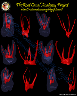
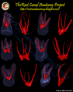
Coronally, the maxillary second molar closely resembles the maxillary first molar, although it is not as square and massive. Access in both teeth can usually be adequately prepared without disturbing the transverse ridge. The second molar is often easier to prepare because of the straight-line access to the orifices. The distinguishing morphologic feature of the maxillary second molar is its three roots grouped close together, and sometimes fused. The parallel root canals are frequently superimposed radiographically. They are usually shorter than the roots of the first molar and not as curved. The three orifices may form a fiat triangle, sometimes almost a straight line. The floor of the chamber is markedly convex, giving a slightly funnel shape to the canal orifices. Occasionally the canals curve into the chamber at a sharp angle to the floor, making it necessary to remove a lip of dentin so the canal can be entered more in a direct line with the canal axis. Complications in access occur when the molar is tipped in distal version. Initial opening with an end-cutting fissure bur is followed by a short-shanked round bur, which is best suited to uncover the pulp chamber and shape the access cavity. Then small hand instruments are used to establish canal continuity and working length. The bulk of the cleaning and shaping may now be accomplished with engine-mounted files on reciprocating handpieces. To enhance radiographic visibility, especially when there is interference with the malar process, a more perpendicular and distal-angular radiograph may be exposed. All caries, leaking restorations, and pulpal calcifications must be removed prior to initiating endodontic treatment. Full occlusal coverage is mandatory to ensure against vertical cuspal or crown-root fracture. Internal reinforcement, when indicated, should be incorporated immediately after endodontic treatment. (Burns RC, Buchanan LS. Tooth Morphology and Access Openings. Part One: The Art of Endodontics in Pathway of Pulp, 6th Ed. p. 148).


Keywords: micro-computed tomography, micro-ct, marco versiani, micro-computer tomography, high resolution x-ray tomography, dental anatomy, root canal anatomy