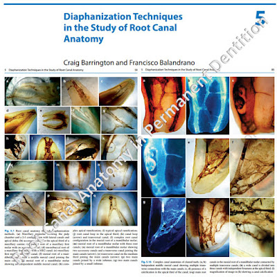
From 2011-2016, images and videos of "The Root Canal Anatomy Project" were developed at the Laboratory of Endodontics of Ribeirao Preto Dental School. From 2016, images were acquired in other educational institutions. They can be freely used for attributed noncommercial educational purposes by educators, scholars, student and clinicians. It means that all material used should include proper attribution and citation (http://rootcanalanatomy.blogspot.com). In such cases, this information should be linked to the image in a manner compatible with such instructional objectives. Unfortunately, because material shared on the RCAP has not been properly cited by several users, from November 2019 a watermark was added to the images and videos. Enjoy!

November 1, 2018

Micro-CT for Dental Applications

The Root Canal Anatomy Project

Dr. Marco Versiani is interviewed by Bruker-microCT company
October, 2018
Dental School of Ribeirao Preto, University of Sao Paulo, Brazil

Original link: https://vimeo.com/297296445
Original link: https://vimeo.com/297957040

October 22, 2018

Webinar: Micro-CT in Endodontics

Endodontic applications of microcomputed tomography 
for studying root canals


Register for free at THIS LINK
Endodontic applications of microcomputed tomography for studying root canals from Bruker Corporation on Vimeo.

Change to a high resolution video at SETTINGS

Overview

In this webinar, researcher in dentistry Dr. Marco Versiani will discuss the applications of micro-CT imaging for 3D root canal studies. Although technological advances in the 20th century have led to a range of techniques for visualizing the human tooth, most require destruction of the samples studied and they also only allow 2D imaging of a 3D structure. Together, these limitations have encouraged the search for new, improved imaging techniques. Over the last decade, non-invasive, non-destructive, high-resolution micro CT technology has become the most important and accurate research tool for the 3D study of root canal anatomy and its influence on treatment/retreatment procedures.

What to expect

The potential and current applications of micro-CT in the analysis of root canal morphology will be described, as well as access, opening, cleaning and shaping procedures and obturation and retreatment protocols. Bruker’s micro-CT systems such as the SkyScan 1174 will also be discussed for analysing various 2D and 3D parameters in dental research.

Key topics

- To explain the current scientific status on micro-CT and the advantages and limitations of using this technology.
- To address the methodological resources provided by the use of micro-CT imaging system in the study of the root canal system and root canal morphology.
- To discuss how further advances of the methodological approaches would progress towards determining the best clinical protocols associated with endodontic procedures.

Who should attend?

The webinar will be of interest to anyone involved in dental and bone research including dentists, biomedical scientists and technicians involved in laboratory studies on the root canal system and anatomy of teeth

August 30, 2018

Video: History of the Studies on Root Canal Anatomy

Historical overview of the studies on root canal anatomy 

by Prof. Marco Versiani


This 47-min video is about the historical aspects of the studies on root canal anatomy, from the classical antiquity to the present days, and it is dedicated to the memory of the pioneers in the endodontic clinical and research fields. If you are a root canal lover, please like this video and share with a colleague.


1080p high quality video is available at settings
Only in English language

May 28, 2018

The Book is Ready!


Book Title: The Root Canal Anatomy in Permanent Dentition
Editors: Marco A. Versiani; Bettina Basrani; Manoel D. Sousa-Neto
Copyright: 2018 Publisher: Springer International Publishing
eBook ISBN: 978-3-319-73444-6 DOI: 10.1007/978-3-319-73444-6
Hardcover ISBN: 978-3-319-73443-9
Edition Number: 1 Number of Pages: XIX, 425
Number of Illustrations and Tables: 26 b/w illustrations, 204 illustrations in colour
Chapters at a glance:


April 7, 2018

Book


Editors: Versiani, Marco A., Basrani, Bettina, Sousa-Neto, Manoel D. (Eds.)

This book describes the most commonly methods used for the study of the internal anatomy of teeth and provides a complete review of the literature concerning the current state of research employing contemporary imaging tools such as micro-CT and CBCT, which offer greater accuracy whether using qualitative or quantitative approaches. In order to facilitate the management of complex anatomic anomalies, specific clinical protocols and valuable practical tips are suggested. In addition, supplementary material consisting in high-quality videos and images of different anatomies obtained using micro-CT technology is made available to the reader. The book was planned and developed in collaboration with an international team comprising world-recognized researchers and experienced clinicians with expertise in the field. It will provide the readers with a thorough understanding of canal morphology and its variations in all groups of teeth, which is a basic prerequisite for the success of endodontic therapy. Due: July, 2018; ISBN 978-3-319-73444-6