From 2011-2016, images and videos of "The Root Canal Anatomy Project" were developed at the Laboratory of Endodontics of Ribeirao Preto Dental School. From 2016, images were acquired in other educational institutions. They can be freely used for attributed noncommercial educational purposes by educators, scholars, student and clinicians. It means that all material used should include proper attribution and citation (http://rootcanalanatomy.blogspot.com). In such cases, this information should be linked to the image in a manner compatible with such instructional objectives. Unfortunately, because material shared on the RCAP has not been properly cited by several users, from November 2019 a watermark was added to the images and videos. Enjoy!

October 31, 2015

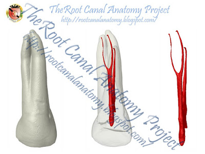
Two-Rooted Maxillary Central Incisor

A review of literature reveals that the anatomy of maxillary central incisors is single-rooted teeth with a single canal in 100% of cases. The prevalence of maxillary central incisors with 2 roots is extremely rare and has never been investigated; only a few clinical case reports have been published (Levin et al. 2015). 


Below there is a similar case report from Levin et al. published in the Journal of Endodontics
This article can be read in full HERE


October 30, 2015

Three-Rooted Maxillary Premolar

The following outstanding clinical case was performed by 
Dr. Konstantinos Kalogeropoulos 
and posted in the Facebook page 'Athens Endo'


Below, the anatomy of a similar three-rooted maxillary premolar is detailed