3D models of root canals textured with computer graphics to resemble pulp tissue
June 29, 2024
May 1, 2024
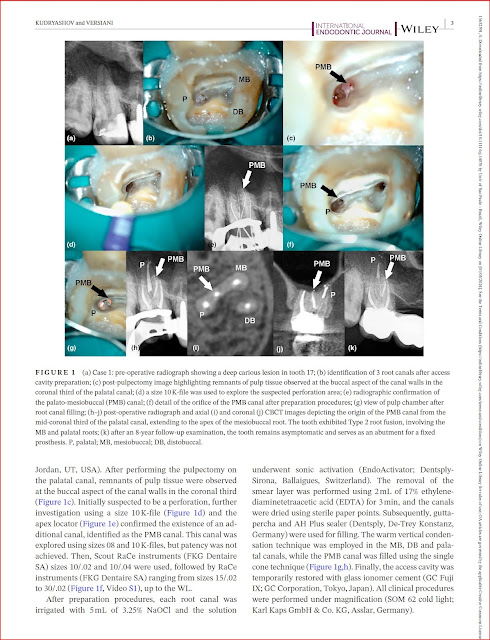
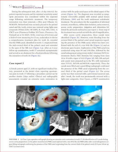
The Palato-Mesiobuccal Canal
Identification and Characterization of a Previously Undiscovered Anatomical Structure in Maxillary Second Molars: The Palato-Mesiobuccal Canal
Marco A Versiani, Tamer Taşdemir, Ali Keleş
Link to the original publication
Anatomical Complexities of the Root Canal System
Anatomical complexities affecting root canal preparation: a narrative review
M A Versiani JNR Martins R Ordinola-Zapata
Link to the original publication
Worldwide Studies: Maxillary Premolars
Worldwide Assessment of the Root and Root Canal Characteristics of Maxillary Premolars - A Multi-center Cone-beam Computed Tomography Cross-sectional Study With Meta-analysis
Jorge N R Martins; Worldwide Anatomy Research Group; Marco A Versiani
Link to the original publication
Worldwide studies: Mandibular Canine
Worldwide Anatomic Characteristics of the Mandibular Canine-A Multicenter Cross-Sectional Study with Meta-Analysis
Jorge N R Martins; Worldwide Anatomy Research Group; Marco A Versiani
Link to the original publication
Pulp Calcification
Micro‑CT assessment of radicular pulp calcifcations in extracted maxillary frst molar teeth
Ali Keleş Cangül Keskin, Marco Versiani
Link to the original publication
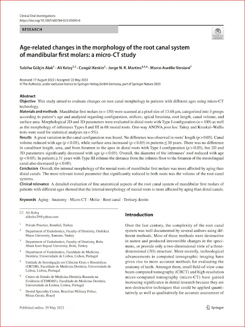
Age‑related changes in the morphology of the root canal system of mandibular frst molars: a micro‑CT study
Sabiha Gülçin Alak, Ali Keleş, Cangül Keskin, Jorge N. R. Martins, Marco Versiani,
Link to the original publication
My Book in Portuguese
Great news! My book on Root Canal Anatomy has just been published in Portuguese. I’m so excited to share this with the Portuguese-speaking dental community!
My images in a Museum in Nuremberg
In October 2022, I was contacted by Dr. des. Susanne Thürigen, a historian of science from the Germanisches Nationalmuseum in Nuremberg, Germany. She expressed interest in using micro-CT images of certain teeth that I had developed to be displayed in the museum's permanent exhibition on crafts and medicine. Now, the images of the root canal anatomy system, which was co-developed by myself, Frank Paqué, and Holm Reuver, are permanently on display in the Spotlight-exhibition Danger Zone in Nuremberg. It's truly an exciting achievement!
#germanishesmuseum
Pulp Fiction 2024 - Poland
Exciting news! From October 5 to 8, 2024, I'll be giving a lecture at the Pulp Fiction Endo Meeting in Krakow, Poland. Can't wait to be part of this amazing event!
Endo Meeting in Portugal 2024
Mark your calendars! I’m thrilled to be lecturing at the IV Meeting of the Portuguese Society of Endodontology from September 27 to 28, 2024. A huge thank you to Dr. Hugo Sousa for the invitation.


