3D models of root canals textured with computer graphics to resemble pulp tissue

Showing posts with label Science & Art. Show all posts
Showing posts with label Science & Art. Show all posts
June 29, 2024


May 1, 2024
My images in a Museum in Nuremberg
In October 2022, I was contacted by Dr. des. Susanne Thürigen, a historian of science from the Germanisches Nationalmuseum in Nuremberg, Germany. She expressed interest in using micro-CT images of certain teeth that I had developed to be displayed in the museum's permanent exhibition on crafts and medicine. Now, the images of the root canal anatomy system, which was co-developed by myself, Frank Paqué, and Holm Reuver, are permanently on display in the Spotlight-exhibition Danger Zone in Nuremberg. It's truly an exciting achievement!
#germanishesmuseum


March 3, 2023
Mandibular First Molar
Root Canal Anatomy Project
Merging Science & Art
Realistic 3D model obtained by micro-CT technology and characterized by using advanced computer design techniques. Watch in HD!
March 2, 2023
Double Maxillary Molar
Root Canal Anatomy Project
Merging Science & Art
Realistic 3D model obtained by micro-CT technology and characterized by using advanced computer design techniques. Watch in HD!
Double Maxillary Molar Teeth
Conjoined teeth, a rare dental anomaly, have been described using various terms including gemination, fusion, double teeth, and twinning. Gemination refers to an attempted division of a single tooth germ, resulting in a tooth with a doubled crown and a single root. Fusion, on the other hand, occurs when two separate tooth germs fuse together, leading to a tooth with a larger crown and two distinct roots. In some cases, it can be challenging to distinguish between the two conditions, which is why some authors prefer to use the term "double teeth" for both. This condition usually affects anterior teeth, but it can also occur in premolars and permanent molars, albeit rarely. In 1963, Tannenbaum and Alling defined gemination as the formation of two teeth from a single follicle, with evidence of an attempt to keep them separate. Pindborg's definition of fusion involves the union of two or more separate developing teeth, which can result in either complete (total/true fusion) or incomplete (partial/late fusion) fusion, depending on the stage of tooth development. Overall, conjoined teeth can present diagnostic challenges, and proper identification and classification are important for appropriate management and treatment.
References
Pindborg JJ. Pathology of the dental hard tissues. Philadelphia: W.B. Saunders; 1970.
Tannenbaum KA, Alling EE. Anomalous tooth development: case report of gemination and twinning. Oral Surg Oral Med Oral Pathol. 1963;16:883-8.
Versiani, M.A., Martins, J.N. & Basrani, B. (2018). 3D visual glossary of terminology in root and root canal anatomy. In M. A. Versiani, B. Basrani & M. D. Sousa Neto (Eds.), The Root Canal Anatomy in Permanent Dentition (1 ed., pp. 391-422). Switzerland: Springer International Publishing.
May 19, 2020


May 17, 2020
Science & Art in the 3D World
Contemporary Approach to Depict the Internal Anatomy of Teeth:
Merging Science & Art in the 3D World



May 16, 2020
New Improvements in Imaging the Anatomy
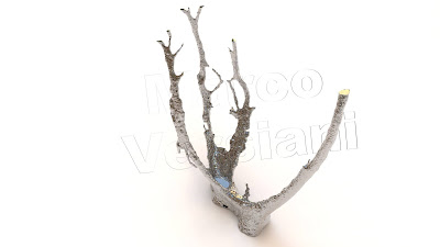
Root Canal Anatomy: The Art that Speaks for Itself
Independent MB2 Canal in a Maxillary First Molar


Subscribe to:
Posts (Atom)